


Page 41 - Concise Pathology for Exam Preparation ( PDFDrive )
P. 41
26    SECTION I  General Pathology


                             •  Chronic arsenical poisoning (raindrop pigmentation)
                             •  Linea nigra (a hyperpigmented line found on the abdomen during pregnancy)
                             •  Café-au-lait spots (neurofibromatosis, Albright syndrome)
                             •  Perioral pigmentation in Peutz–Jeghers syndrome
                             •  Melanocytic tumours/nevi
                             •  Dermatopathic lymphadenitis
                           •  Hypopigmentation:
                             •  Albinism
                             •  Vitiligo
                             •  Leprosy
                             •  Post-inflammatory scarring
                             •  Radiation dermatitis
                     Staining characteristics of melanin:
                     Can  be  bleached  with  hydrogen  peroxide  and  stained  with  Masson–Fontana argentaffin
                     stain; this forms the basis of differentiation of melanin from melanin lookalikes, eg, homo-
                     gentisic acid seen in alkaptonuria and carbon seen in anthracosis.
                      (b)  Lipofuscin
                           (i)  Lipid-derived wear and tear pigment (associated with atrophied cells of old age
                             and wasting)
                          (ii)  Derived from the Latin word ‘fuscus’, meaning brown
                           (iii)  Sometimes called ‘residual bodies’ (collection of indigestible material in the lyso-
                             somes after intracellular lipid peroxidation)
                          (iv)  Yellow-brown, granular, intracytoplasmic (perinuclear in location)
                          (v)  Seen in myocardium, hepatocytes, Leydig cells and neurons
                     Staining characteristics of lipofuscin:
                     •  Acid fast (AFB positive)
                     •  Autofluorescent
                     •  Stains positive with fat stains
                     •  Reduces ferricyanide to ferrocyanide (Schmorl reaction)
                      (c)  Haemosiderin
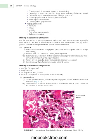
                          (i)  Golden-yellow to brown, crystalline granular pigment, which stains with Prussian
                            blue stain (Fig. 1.17).
                           (ii)  Haemosiderosis is defined as the presence of stainable iron in tissue. Based on
                            distribution, it may be classified as


                                                                                 Haemosiderin
                                                                                 deposits


                           FIGURE 1.17.  Haemosiderin deposits in the lining of a bone cyst (H&E; 4003).


                                  mebooksfree.com

