


Page 38 - Concise Pathology for Exam Preparation ( PDFDrive )
P. 38
1  Cell Injury and Cell Death  23


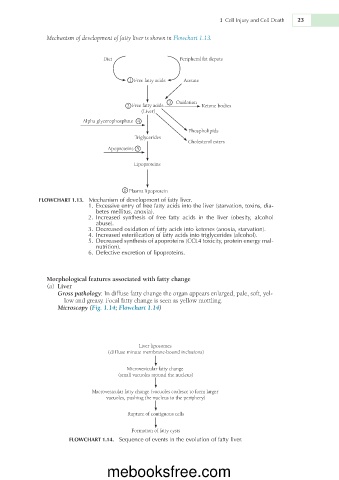
               Mechanism of development of fatty liver is shown in Flowchart 1.13.


                                 Diet                   Peripheral fat depots


                                         1  Free fatty acids  Acetate


                                                     3  Oxidation
                                        2  Free fatty acids    Ketone bodies
                                             (Liver)
                           Alpha glycerophosphate 4
                                                           Phospholipids
                                          Triglycerides
                                                           Cholesterol esters
                                  Apoproteins 5

                                          Lipoproteins


                                       6  Plasma lipoprotein
             FLOWCHART 1.13.  Mechanism of development of fatty liver.
                            1.  Excessive entry of free fatty acids into the liver (starvation, toxins, dia-
                               betes mellitus, anoxia).
                            2.  Increased  synthesis  of  free  fatty  acids  in  the  liver  (obesity,  alcohol
                               abuse).
                            3.  Decreased oxidation of fatty acids into ketones (anoxia, starvation).
                            4.  Increased esterification of fatty acids into triglycerides (alcohol).
                            5.  Decreased synthesis of apoproteins (CCL4 toxicity, protein energy mal-
                               nutrition).
                            6.  Defective excretion of lipoproteins.


               Morphological features associated with fatty change
                (a)  Liver
                   Gross pathology: In diffuse fatty change the organ appears enlarged, pale, soft, yel-
                     low and greasy. Focal fatty change is seen as yellow mottling.
                   Microscopy (Fig. 1.14; Flowchart 1.14)


                                            Liver liposomes
                                  (diffuse minute membrane-bound inclusions)

                                        Microvesicular fatty change
                                     (small vacuoles around the nucleus)

                             Macrovesicular fatty change (vacuoles coalesce to form larger
                                  vacuoles, pushing the nucleus to the periphery)

                                        Rupture of contiguous cells

                                          Formation of fatty cysts
                      FLOWCHART 1.14.  Sequence of events in the evolution of fatty liver.


                                  mebooksfree.com

