
Page 414 - Concise Pathology for Exam Preparation ( PDFDrive )
P. 414
14 The Oral Cavity and Gastrointestinal Tract 399
• Increases production of proinflammatory cytokines, eg, IL-1, IL-6, TNF and IL-8
(chemotactic to neutrophils).
• May cause thrombotic occlusion causing ischaemia.
• Epithelial injury is induced by VacA (a vacuolating toxin) regulated by CagA (cytotoxin-
associated gene).
H. pylori is associated with duodenal ulcer in 80–90% patients and gastric ulcer
in 60% patients. Virulence of the infecting strain determines development of peptic ulcer
in an individual infected with H. pylori.
Gross Morphology
• Small, round-to-oval, sharply punched-out ulcers, varying in size between 2 and 4 cm.
• Straight wall, mucosa may overhang the base.
• Base—clean and smooth (due to peptic digestion)
Microscopic Features
In an active ulcer, four distinct zones are appreciated (Askanazy zones; Fig. 14.6):
1. Zone of fibrinoid necrosis
2. Zone of nonspecific inflammatory infiltrate (predominantly neutrophils)
3. Zone of granulation tissue (proliferating blood vessels, fibroblasts and mononuclear
cells)
4. Zone of fibrosis (collagenous or fibrous scar formation)
Complications
• Bleeding: Most frequent complication (seen in 15–20% of the patients); may be life
threatening, and sometimes, is the first indication of presence of peptic ulcer.
• Perforation: Life-threatening late presentation (responsible for two-third of the deaths
due to ulcers); may result in acute peritonitis and subphrenic abscess; may involve
adjacent organs.
• Obstruction: Occurs due to oedema or scarring
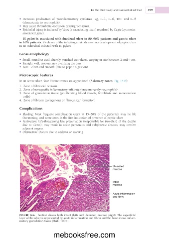
Ulcerated
mucosa
Intact
mucosa
Acute inflammation
and fibrin
FIGURE 14.6. Section shows both intact (left) and ulcerated mucosa (right). The superficial
layer of the ulcer is represented by acute inflammation and fibrin and the base shows inflam-
matory granulation tissue (H&E; 1003).
mebooksfree.com