
Page 412 - Concise Pathology for Exam Preparation ( PDFDrive )
P. 412
14 The Oral Cavity and Gastrointestinal Tract 397
• Autoimmunity: Presence of autoantibodies, to gastric parietal cells, mainly to the acid-
producing enzyme H1/K1-ATPase leading to loss of both acid-producing and intrinsic
factor-producing cells. The gastric corpus (body) undergoes progressive atrophy. Its sequelae
include development of pernicious anaemia, adenocarcinoma and gastric carcinoid.
• Toxic substances: Alcohol intake and tobacco smoking
• Iatrogenic causes: Postsurgical (antrectomy and gastroenterostomy)
• Radiation exposure: Radiation-induced gastritis is an infrequent cause of gastrointestinal
bleeding.
• Infectious granulomatous gastritis: Granulomatous gastritis is a rare entity caused by
organisms like M. tuberculosis and fungi usually in patients who are immunosuppressed.
• Chronic reactive chemical gastropathy: Gastritis may result from long-term intake of
aspirin or NSAIDs. It also develops when bile-containing intestinal contents reflux into
the stomach.
• Others: Amyloidosis and graft versus host reactions
Gross Morphology
Mucosa reddened, coarse with thick rugal folds in early, and thinned with flattened rugal
folds in long-standing disease
Microscopic Features
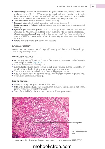
• Lamina propria is infiltrated by chronic inflammatory infiltrate composed of lympho-
cytes and plasma cells. (Fig. 14.5)
• Intestinal metaplasia is frequently seen
• In long-standing disease due to H. pylori as well as autoimmune gastritis, there is loss or
atrophy of parietal cells, leading to hypochlorhydria or achlorhydria
• This, in turn, may induce G-cell hyperplasia and hypergastrinaemia.
• H. pylori, if present, lies in the superficial mucosal layer among the microvilli of epithelial cells.
• Occasionally, dysplasia may develop.
Clinical Features
• Nausea, vomiting and upper abdominal discomfort
• Mild form: Hypochlorhydria (not achlorhydria), pernicious anaemia absent and serum
gastrin level normal or slightly increased
• Severe form: Achlorhydria, pernicious anaemia and hypergastrinaemia
Gastric glands
Chronic inflammatory
cells
FIGURE 14.5. Gastric mucosa showing chronic nonspecific inflammation (H&E; 2003).
mebooksfree.com