
Page 416 - Concise Pathology for Exam Preparation ( PDFDrive )
P. 416
14 The Oral Cavity and Gastrointestinal Tract 401
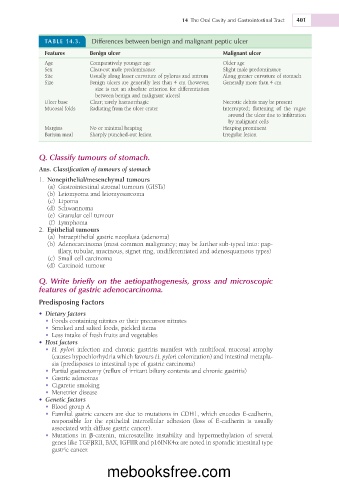
TABLE 14.3. Differences between benign and malignant peptic ulcer
Features Benign ulcer Malignant ulcer
Age Comparatively younger age Older age
Sex Clear-cut male predominance Slight male predominance
Site Usually along lesser curvature of pylorus and antrum Along greater curvature of stomach
Size Benign ulcers are generally less than 4 cm (however, Generally more than 4 cm
size is not an absolute criterion for differentiation
between benign and malignant ulcers)
Ulcer base Clear; rarely haemorrhagic Necrotic debris may be present
Mucosal folds Radiating from the ulcer crater Interrupted; flattening of the rugae
around the ulcer due to infiltration
by malignant cells
Margins No or minimal heaping Heaping prominent
Barium meal Sharply punched-out lesion Irregular lesion
Q. Classify tumours of stomach.
Ans. Classification of tumours of stomach
1. Nonepithelial/mesenchymal tumours
(a) Gastrointestinal stromal tumours (GISTs)
(b) Leiomyoma and leiomyosarcoma
(c) Lipoma
(d) Schwannoma
(e) Granular cell tumour
(f) Lymphoma
2. Epithelial tumours
(a) Intraepithelial gastric neoplasia (adenoma)
(b) Adenocarcinoma (most common malignancy; may be further sub-typed into: pap-
illary, tubular, mucinous, signet ring, undifferentiated and adenosquamous types)
(c) Small cell carcinoma
(d) Carcinoid tumour
Q. Write briefly on the aetiopathogenesis, gross and microscopic
features of gastric adenocarcinoma.
Predisposing Factors
• Dietary factors
• Foods containing nitrites or their precursor nitrates
• Smoked and salted foods, pickled items
• Less intake of fresh fruits and vegetables
• Host factors
• H. pylori infection and chronic gastritis manifest with multifocal mucosal atrophy
(causes hypochlorhydria which favours H. pylori colonization) and intestinal metapla-
sia (predisposes to intestinal type of gastric carcinoma)
• Partial gastrectomy (reflux of irritant biliary contents and chronic gastritis)
• Gastric adenomas
• Cigarette smoking
• Menetrier disease
• Genetic factors
• Blood group A
• Familial gastric cancers are due to mutations in CDH1, which encodes E-cadherin,
responsible for the epithelial intercellular adhesion (loss of E-cadherin is usually
associated with diffuse gastric cancer).
• Mutations in b-catenin, microsatellite instability and hypermethylation of several
genes like TGFbRII, BAX, IGFIIR and p16INK4a are noted in sporadic intestinal type
gastric cancer.
mebooksfree.com