
Page 492 - Concise Pathology for Exam Preparation ( PDFDrive )
P. 492
16 Diseases of the Kidney and Lower Urinary Tract 477
• Usually self-limiting; in the presence of predisposing conditions may become recur-
rent or chronic.
2. Chronic pyelonephritis (CPN) and reflux nephropathy
Morphological entity in which interstitial inflammation and scarring of renal paren-
chyma is associated with scarring and deformity of the pelvocalyceal system.
Types:
(a) Chronic obstructive pyelonephritis
- Recurrent infections occurring in a background of obstruction which lead to
repeated inflammation and scarring.
- The disease can be bilateral as in congenital anomalies of the urethra (posterior
urethral valves) or unilateral as in calculi and unilateral obstructive lesions.
(b) Chronic reflux–associated pyelonephritis
This more common form of CPN results from the superimposition of UTI on con-
genital vesicoureteral reflux and intrarenal reflux. May be unilateral or bilateral.
Gross Morphology:
• May be unilateral or bilateral, patchy or diffuse.
• Coarse, discrete corticomedullary scars are seen corresponding to the overlying
blunted or dilated calyces.
• Asymmetrical pelvocalyceal scarring leads to blunting of papillae and deformity of calyces.
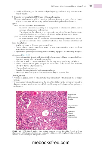
Microscopy (Fig. 16.9):
• Uneven interstitial fibrosis with interstitial inflammatory infiltrate composed of lym-
phocytes, plasma cells and rarely neutrophils.
• Dilatation as well as contraction of tubules showing atrophy of lining epithelium.
• Dilated tubules contain pink PAS-positive casts called ‘colloid casts’ that resemble
colloid in thyroid (thyroidization).
• Fibrosis of calyceal mucosa.
• Vascular changes similar to benign arteriosclerosis.
• Late stages may show glomerulosclerosis secondary to nephron loss.
Clinical features:
• Presents as gradual onset of renal insufficiency (azotaemia); often noticed due to hyper-
tension.
• Ultrasonography is used to determine the size of the kidney and a pyelogram is used to
show the asymmetrical contraction of kidneys, blunting and deformity of the pelvocaly-
ceal system.
Glomerulus
with
periglomerular
tibrasis
Thyroidization
of tubules
Interstitial
tibrasis
Interstitial
inflammation
FIGURE 16.9. H&E-stained section from kidney showing uneven interstitial fibrosis with an
inflammatory infiltrate and dilatation and contraction of tubules with atrophy of lining epithelium.
Dilated tubules contain pink PAS-positive casts.
mebooksfree.com