
Page 559 - Concise Pathology for Exam Preparation ( PDFDrive )
P. 559
544 SECTION II Diseases of Organ Systems
Q. Write briefly on the pathogenesis and clinicomorphological
features of adenomas of thyroid.
Ans. Adenomas are discrete, solitary masses derived from the follicular epithelium (thus,
also called follicular adenomas). Hormone production in functional adenomas (also called
toxic adenomas) is independent of TSH stimulation. This is labelled thyroid autonomy.
Majority of the adenomas are nonfunctional (take up less iodine than normal thyroid tissue
and appear as cold nodules). Functioning adenomas appear as hot nodules.
Pathogenesis of Nonfunctioning Adenomas
Nonfunctioning adenomas may have any of the following genetic alterations:
1. Mutations in RAS proto-oncogene
2. Phosphatidylinositol-3-kinase subunit abnormalities
3. PAX8-PPARG fusion gene alterations
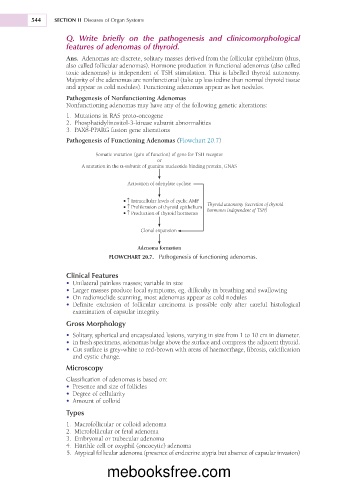
Pathogenesis of Functioning Adenomas (Flowchart 20.7)
Somatic mutation (gain of function) of gene for TSH receptor
or
A mutation in the α-subunit of guanine nucleotide binding protein, GNAS
Activation of adenylate cyclase
• ↑ Intracellular levels of cyclic AMP Thyroid autonomy (secretion of thyroid
• ↑ Proliferation of thyroid epithelium hormones independent of TSH)
• ↑ Production of thyroid hormones
Clonal expansion
Adenoma formation
FLOWCHART 20.7. Pathogenesis of functioning adenomas.
Clinical Features
• Unilateral painless masses; variable in size
• Larger masses produce local symptoms, eg, difficulty in breathing and swallowing
• On radionuclide scanning, most adenomas appear as cold nodules
• Definite exclusion of follicular carcinoma is possible only after careful histological
examination of capsular integrity.
Gross Morphology
• Solitary, spherical and encapsulated lesions, varying in size from 1 to 10 cm in diameter.
• In fresh specimens, adenomas bulge above the surface and compress the adjacent thyroid.
• Cut surface is grey-white to red-brown with areas of haemorrhage, fibrosis, calcification
and cystic change.
Microscopy
Classification of adenomas is based on:
• Presence and size of follicles
• Degree of cellularity
• Amount of colloid
Types
1. Macrofollicular or colloid adenoma
2. Microfollicular or fetal adenoma
3. Embryonal or trabecular adenoma
4. Hürthle cell or oxyphil (oncocytic) adenoma
5. Atypical follicular adenoma (presence of endocrine atypia but absence of capsular invasion)
mebooksfree.com