
Page 603 - Concise Pathology for Exam Preparation ( PDFDrive )
P. 603
588 SECTION II Diseases of Organ Systems
• The cyst expands the bone, causing thinning of the overlying cortex.
• Pathogenesis is unknown.
• SBC may remain asymptomatic or present with pain and pathological fracture.
Gross pathology
Generally unilocular with smooth inner lining; filled with yellow or amber coloured
fluid.
Microscopy
• Cyst wall consists of thin collagenous tissue having scattered osteoclastic giant cells
and newly formed reactive bony trabeculae.
• Fracture may alter the appearance with secondary haemorrhage, haemosiderin
deposits and macrophages in the cyst wall.
2. Aneurysmal bone cyst (ABC)
• ABC is an expanding osteolytic lesion filled with blood (aneurysm 5 dilatation).
• Common in young patients under 30 years of age.
• Most frequently involved is metaphysis of long bones or the vertebral column.
X-Ray
Characteristic ballooned-out, expansile lesion located underneath the periosteum
Pathogenesis
Not clear; probably arises from persistent alteration in the local haemodynamics
Clinical features
Enlarges over a period of years to produce pain, tenderness and sometimes pathological
fracture
Gross pathology
Seen as a large haemorrhagic mass covered over by thinned out reactive bone
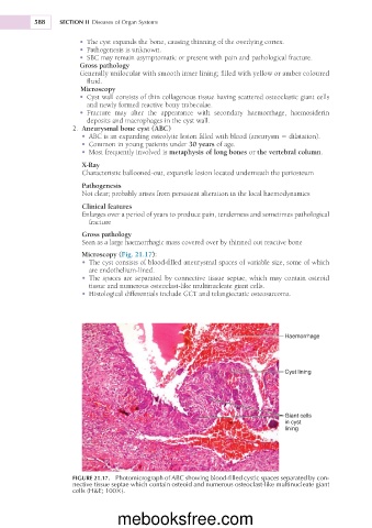
Microscopy (Fig. 21.17):
• The cyst consists of blood-filled aneurysmal spaces of variable size, some of which
are endothelium-lined.
• The spaces are separated by connective tissue septae, which may contain osteoid
tissue and numerous osteoclast-like multinucleate giant cells.
• Histological differentials include GCT and telangiectatic osteosarcoma.
Haemorrhage
Cyst lining
Giant cells
in cyst
lining
FIGURE 21.17. Photomicrograph of ABC showing blood-filled cystic spaces separated by con-
nective tissue septae which contain osteoid and numerous osteoclast-like multinucleate giant
cells (H&E; 1003).
mebooksfree.com