


Page 61 - Concise Pathology for Exam Preparation ( PDFDrive )
P. 61
46    SECTION I  General Pathology


                        Manifestations of SIRS include
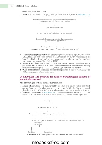
                       1.  Fever: The mechanism underlying development of fever is depicted in Flowchart 2.12.

                                   Bacterial products (exogenous pyrogens) act on leukocytes to release
                                          cytokines IL-1 and TNF (endogenous pyrogens)


                                                 Increased cyclooxygenase

                                       Increased conversion of arachidonic acid to prostaglandins


                                      Prostaglandin E stimulates production of neurotransmitters
                                              (eg, cyclic AMP) in hypothalamus


                                           Resetting of the temperature at a higher level
                                FLOWCHART 2.12.  Mechanism of development of fever in SIRS.

                       2.  Release of acute phase proteins: Acute phase proteins/reactants, eg, C reactive protein
                        (CRP), fibrinogen and serum amyloid A (SAA) protein, are mostly synthesized in the
                        liver. They bind to the cell wall (act as opsonins) and complement, and their synthesis
                        is regulated by cytokines IL-1, IL-6 and TNF.
                       3.  Leukocytosis: Accelerated release of cells from the bone marrow post-mitotic reserve
                        pool with a shift to left (due to IL-1 and TNF) is common in inflammation and this may
                        result in counts as high as 40,000–100,000 cells/µL (leukaemoid reaction).
                       4.  Other manifestations: Increased pulse and blood pressure, decreased sweating, rigours,
                        chills, anorexia, somnolence and malaise

                     Q. Enumerate and describe the various morphological patterns of
                     acute inflammation?
                     Ans. Morphologic patterns of acute inflammation:
                       1.  Serous inflammation: It is characterized by collection of a watery, protein-poor fluid;
                        derived  from  either  the  plasma  or  secretions  of  mesothelial  cells  (lining  peritoneal,
                        pleural and pericardial cavities). It is usually associated with burns, viral infections, etc.
                      2.  Fibrinous inflammation (Flowchart 2.13): Examples of fibrinous inflammation include
                        ‘bread and butter’ pericarditis seen in acute rheumatic fever and fibrinous pleuritis.


                                              Severe injury


                                        Increased vascular permeability


                               Larger molecules like fibrinogen pass the vascular barrier


                                  •  Fibrin is formed and deposited in the extracellular space (seen in meninges and
                                     pericardium)
                                  •  May undergo


                              Resolution      Organization
                             FLOWCHART 2.13.  Pathogenesis and outcomes of fibrinous inflammation.


                                  mebooksfree.com

