


Page 99 - Concise Pathology for Exam Preparation ( PDFDrive )
P. 99
84    SECTION I    General Pathology

                     Q.   Differentiate between red and white infarcts.

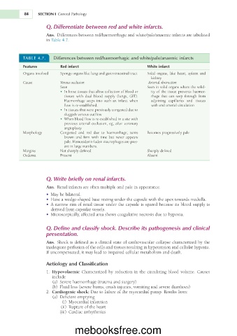
                     Ans.    Differences   between   red/haemorrhagic   and   white/pale/anaemic   infarcts   are   tabulated
                     in  Table   4.7.


           TABLE   4.7.   Differences between red/haemorrhagic and white/pale/anaemic infarcts

           Features         Red infarct	�                         White infarct


           Organs   involved   Spongy   organs   like   lung   and   gastrointestinal   tract   Solid    organs,  like  heart,  spleen  and
                                                                    kidney
           Cause            Venous occlusion                       Arterial obstruction
                                                                        solid
                            Seen                                  Seen  in    organs   where   the   solid-
                              •  In   loose   tissues   that   allow   collection  of        ity  of
                                                         blood  or
                                                                         the  tissue  prevents  haemor-


                             tissues    with  dual  blood  supply  (lungs,  GIT).     rhage    that  can  seep  through  from


                             Haemorrhage   seeps  into  such  an  infarct  when     adjoining    capillaries  and  tissues


                             flow      is  re-established.          with   end   arterial   circulation
                              •  In   tissues   that   were   previously   congested   due  to

                             sluggish   venous   outflow
                              •  When   blood   flow  is        site   with
                                            re-established  in  a


                             previous    arterial  occlusion,  eg,  after  coronary
                             angioplasty

           Morphology       Congested    and  red  due  to  haemorrhage;  turns     Becomes   progressively   pale


                             brown    and  firm  with  time  but  never  appears

                             pale.   Hemosiderin-laden   macrophages   are   pres-
                             ent  in    numbers.
                                  large
           Margins          Not   sharply   defined               Sharply   defined
           Oedema           Present                               Absent
                     Q.   Write briefly on renal infarcts.
                     Ans.    Renal   infarcts   are   often   multiple   and   pale     appearance.
                                                            in
                           be
                        •	�May     bilateral.
                        •	�Have     wedge-shaped   base   resting   under   the   capsule   with   the   apex   towards   medulla.
                            a

                         narrow  rim
                                                                                            is

                                                               is


                                   of


                     •	� A            renal  tissue  under  the  capsule     spared  because  its  blood  supply
                       derived   from   capsular   vessels.
                        •	�Microscopically,   affected   area   shows   coagulative   necrosis   due     hypoxia.
                                                                         to
                     Q.   Define and classify shock. Describe its pathogenesis and clinical

                     presentation.
                                                       of

                                is
                                         as


                                           a
                     Ans.    Shock     defined       clinical  state     cardiovascular  collapse  characterized  by  the


                     inadequate   perfusion     the   cells   and   tissues   resulting     hypotension   and   cellular   hypoxia.
                                       of
                                                                in
                                     it
                         If  uncompensated,     may   lead     impaired   cellular   metabolism   and   death.
                                              to
                     Aetiology and Classification


                                                            in


                     1.                                       the  circulating  blood  volume.  Causes
                        	 Hypovolaemic  Characterized  by  reduction
                        include
                        (a)
                            Severe   haemorrhage   (trauma   and   surgery)
                            Fluid
                        (b)      loss   (severe   burns,   crush   injuries,   vomiting   and   severe   diarrhoea)
                     2.
                        	 Cardiogenic shock: Due     failure     the   myocardial   pump.   Results   from:
                                                     of
                                             to
                            Deficient   emptying
                        (a)

                             (i)  Myocardial   infarction

                            (ii)  Rupture     the   heart
                                      of
                             (iii)  Cardiac   arrhythmias

                                  mebooksfree.com

