
Page 1328 - Hematology_ Basic Principles and Practice ( PDFDrive )
P. 1328
1174 Part VII Hematologic Malignancies
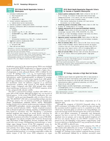
TABLE 2016 World Health Organization Variants of TABLE 2016 World Health Organization Diagnostic Criteria
72.3 Mastocytosis 72.4 for Variants of (Systemic) Mastocytosis
1. Cutaneous mastocytosis (CM) 1. Indolent systemic mastocytosis (ISM): Meets criteria for SM. No “B”
a. Maculopapular CM or “C” findings. No evidence of associated clonal hematologic
b. Diffuse CM malignancy/disorder. In this variant, the mast cell burden is usually
c. Mastocytoma of skin low; skin lesions are almost invariably present.
2. Indolent systemic mastocytosis (ISM) • Bone marrow mastocytosis: As above, with bone marrow
a. Isolated bone marrow mastocytosis involvement, but no skin lesions.
3. Smoldering systemic mastocytosis (SSM) 2. Smoldering systemic mastocytosis (SSM): Meets criteria for SM. Two
4. Systemic mastocytosis with an associated hematologic neoplasm or more “B” findings and no “C” findings.
(SM-AHN)* 3. Systemic mastocytosis with an associated hematologic neoplasm
a. SM-MDS (SM-AHN)*: Meets criteria for SM and criteria for an associated
b. SM-MPN (e.g. PV, ET, MF, CML) hematologic neoplasm (MDS, MPN, MDS/MPN, CEL, AML,
c. SM-MDS/MPN (e.g. CMML, MDS/MPN-unclassified) lymphoma, or other hematologic neoplasm that meets the criteria
d. SM-CEL for a distinct entity in the WHO classification).
e. SM-AML 3. Aggressive systemic mastocytosis (ASM): Meets criteria for SM. One
f. SM-lymphoid neoplasm (e.g. NHL, CLL, multiple myeloma) or more “C” findings. No associated clonal hematologic neoplasm.
5. Aggressive systemic mastocytosis (ASM) No evidence of mast cell leukemia.
6. Mast cell leukemia (MCL) 4. Mast cell leukemia (MCL): Meets criteria for SM. Bone marrow biopsy
a. Aleukemic MCL shows diffuse infiltration, usually interstitial pattern, by atypical,
7. Mast cell sarcoma (MCS) immature mast cells. Bone marrow aspirate smears show 20% or
*SM-AHN is a new term that may be used in lieu of, or interchangeably with more mast cells. Cases in which <10% of circulating WBCs are
the previously used term, SM-AHNMD (systemic mastocytosis with an mast cells are referred to as ‘aleukemic mast cell leukemia’.
associated non-mast cell lineage disease). 5. Mast cell sarcoma (MCS): Unifocal mast cell tumor. No evidence of
AML, Acute myeloid leukemia; CEL, chronic eosinophilic leukemia; CLL, SM. No skin lesions. Destructive growth pattern. High-grade
chronic lymphocytic leukemia; CML, chronic myeloid leukemia; CMML, chronic
myelomonocytic leukemia; ET, essential thrombocythemia; MDS, cytology.
myelodysplastic syndrome; MF, myelofibrosis; MPN, myeloproliferative *SM-AHN is a new term that may be used in lieu of, or interchangeably with
neoplasm; NHL, non–Hodgkin lymphoma; PV, polycythemia vera. the previously used term, SM-AHNMD (systemic mastocytosis with an
associated non-mast cell lineage disease).
AML, Acute myeloid leukemia; CEL, chronic eosinophiic leukemia; MDS,
myelodysplastic syndrome; MPN, myeloproliferative neoplasm; WBC, white
blood cell; WHO, World Health Organization.
classification proposed by the consensus group, SSM is now included
in the revised 2016 WHO classification as a separate variant of SM,
which is meaningful because these patients exhibit a higher chance
of progression to more advanced disease. SSM is defined by two TABLE
or more “B” findings (Table 72.5), e.g., (1) hepatomegaly without 72.5 “B” Findings: Indication of High Mast Cell Burden
impairment of liver function, and/or palpable splenomegaly without
hypersplenism, and/or lymphadenopathy on palpation or imaging; 1. Infiltration grade (mast cells) greater than 30% in bone marrow in
(2) BM MC burden >30% and serum tryptase level >200 ng/mL; histology and serum total tryptase levels greater than 200 ng/mL
and (3) signs of dysplasia or myeloproliferation, in non-MC lineage(s), 2. Hypercellular marrow with loss of fat cells, discrete signs of
but insufficient criteria for definitive diagnosis of an additional dysmyelopoiesis without substantial cytopenias, and without WHO
hematopoietic neoplasm, with normal or only slightly abnormal criteria for an MDS or MPN
blood counts. Compared with ISM, SSM patients often exhibit 3. Organomegaly: palpable hepatomegaly, splenomegaly, or
clonal multilineage involvement of the KIT D816V mutation, which lymphadenopathy (on CT or ultrasound) greater than 2 cm without
is also a prognostically relevant variable. impaired organ function
SM with an associated hematologic neoplasm (SM-AHN) com- MDS, Myelodysplastic syndrome; MPN, myeloproliferative neoplasm;
prises 30% of SM variants, although this figure may vary because of WHO, World Health Organization.
referral patterns. In the revised 2016 WHO classification, the term
“SM-AHN” can be used interchangeably with or in lieu of the prior
term “SM-AHNMD” (SM with an associated hematologic non-mast
cell lineage disease). The vast majority of AHNs are myeloid neo- dysfunction, especially when both disease components are of an
plasms: myelodysplastic syndrome (MDS), MPNs, MDS/MPN aggressive type.
overlap disorders such as chronic myelomonocytic leukemia (CMML) Although the prognosis of SM-AHN frequently relates to the
or MDS/MPN-unclassified, chronic eosinophilic leukemia (CEL), AHN component, the burden of SM as well as the type and stage of
12
and acute myeloid leukemia (AML). Rarely, lymphoid neoplasms the associated myeloid neoplasm need to be considered on an indi-
such as chronic lymphocytic leukemia, myeloma, or lymphomas have vidual basis. Treatment plans are tailored to the histopathologic and
been found in association with SM. Identifying a concomitant molecular findings and the clinical sequelae that are felt to be attribut-
myeloid neoplasm may depend on several factors, including the able to each disease component. A commonly cited therapeutic
expertise of the evaluating pathologist, and whether one disease is approach has been to treat the SM component as if the myeloid
masked by the presence of another. For example, in cases of SM-AML, neoplasm were not present, and to treat the myeloid neoplasm as if
MC aggregates may only be unmasked after induction chemotherapy SM were not present. Because the KIT D816V mutation may be
with achievement of BM hypoplasia, because neoplastic MCs may present in the cells belonging to both disease compartments, small
persist after such therapy. Distinguishing nonhematologic or hema- molecule inhibitors of dysregulated KIT may provide benefit for both
tologic organ damage because of the SM component versus the the SM and AHN in selected cases.
associated myeloid disease can be very difficult, if not impossible, in Aggressive systemic mastocytosis (ASM) comprises 5%–10% of
some patients. Even when a biopsy of the involved extramedullary SM variants and is defined by one or more “C findings” (Table 72.6)
organ is analyzed to elucidate the burden of neoplastic MCs versus reflecting organ dysfunction because of neoplastic MC infiltrates.
associated myeloid neoplasm, it is sometimes impossible to define the Examples of C findings include marked cytopenias because of exten-
relative impact of the SM versus AHN component on organ sive BM involvement (defined by an absolute neutrophil count