
Page 1422 - Hematology_ Basic Principles and Practice ( PDFDrive )
P. 1422
1268 Part VII Hematologic Malignancies
1,7
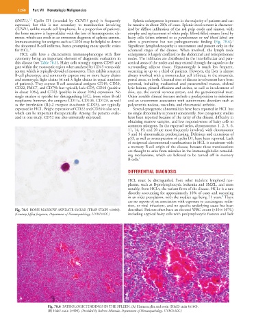
(SMZL). Cyclin D1 (encoded by CCND1 gene) is frequently Splenic enlargement is present in the majority of patients and can
expressed, but this is not secondary to translocation involving be massive in about 20% of cases. Splenic involvement is character-
CCND1, unlike mantle cell lymphoma. In a proportion of patients, ized by diffuse infiltration of the red pulp cords and sinuses, with
the bone marrow is hypocellular with the loss of hematopoietic ele- atrophy and replacement of white pulp. Blood-filled sinuses lined by
ments, which can result in an erroneous diagnosis of aplastic anemia. hairy cells (often referred to as pseudosinuses or red blood lakes) are
1
Immunostaining for antigens such as CD20 may be helpful to detect often a prominent but not pathognomonic finding (Fig. 78.6).
the abnormal B-cell infiltrate, hence prompting more specific stains Significant lymphadenopathy is uncommon and present only in the
for HCL. 1 advanced stages of the disease. When involved, the lymph node
HCL cells have a characteristic immunophenotype with flow enlargement is largely confined to the abdominal and retroperitoneal
cytometry being an important element of diagnostic evaluation in nodes. The infiltrates are distributed in the interfollicular and para-
this disease (see Table 78.1). Hairy cells strongly express CD45 and cortical areas of the nodes and may extend through the capsule to the
gate within the monocytic region when analyzed by CD45 versus side surrounding adipose tissue. Hepatomegaly is much less frequent,
scatter, which is typically devoid of monocytes. They exhibit a mature occurring in up to a third of patients. However, the liver is almost
B-cell phenotype and commonly express one or more heavy chains always involved with a mononuclear cell infiltrate in the sinusoids,
and monotypic light chains (κ and λ light chains in equal numbers portal areas, or both. Unusual sites of disease involvement have been
of patients). They express B-cell associated antigens CD19, CD20, reported, including mediastinal and paravertebral masses, skeletal
CD22, FMC7, and CD79b but typically lack CD5, CD10 (positive lytic lesions, pleural effusions and ascites, as well as involvement of
in about 10%), and CD23 (positive in about 20%) expression. No skin, eye, the central nervous system, and the gastrointestinal tract.
single marker is specific for distinguishing HCL from other B-cell Other notable clinical features include a predisposition to infections
neoplasms; however, the antigens CD11c, CD103, CD123, as well and an uncommon association with autoimmune disorders such as
as the interleukin (IL)-2 receptor α-subunit (CD25), are typically polyarteritis nodosa, vasculitis, and rheumatoid arthritis.
expressed in HCL. Bright expression of CD22 and CD20 is also seen, Several cytogenetic abnormalities have been reported in HCL but
which can be important therapeutically. Among the patients evalu- no single abnormality is present consistently. Few cytogenetic studies
ated in one study, CD52 was also universally expressed. have been reported because of the rarity of the disease, difficulty in
obtaining marrow samples, and low responsiveness of hairy cells to
common mitogens. In the reported series, chromosomes 1, 2, 5, 6,
11, 14, 19, and 20 are most frequently involved, with chromosome
5 and 14 abnormalities predominating. Deletions and mutations of
p53, as well as overexpression of cyclin D1, have been reported. Lack
of reciprocal chromosomal translocations in HCL is consistent with
a memory B-cell origin of the disease, because these translocations
are thought to arise from mistakes in the immunoglobulin remodel-
ing mechanisms, which are believed to be turned off in memory
B cells. 2
DIFFERENTIAL DIAGNOSIS
HCL must be distinguished from other indolent lymphoid neo-
plasms, such as B-prolymphocytic leukemia and SMZL, and most
notably, from HCLv, the variant form of the disease. HCLv is a rare
disorder accounting for approximately 10% of cases and occurring
8
in an older population, with the median age being 71 years. There
are no reports of an association with exposure to carcinogens, radia-
tion, or viral infections, and no specific underlying cause has been
9
Fig. 78.5 BONE MARROW ASPIRATE SMEAR (TRAP STAIN ×1000). described. Patients often have an elevated WBC count (>10 × 10 /L)
(Courtesy Jeffrey Jorgensen, Department of Hematopathology, UTMDACC.) including atypical hairy cells with prolymphocytic features and lack
A B
Fig. 78.6 PATHOLOGIC FINDINGS IN THE SPLEEN. (A) Hematoxylin and eosin (H&E) stain (×100).
(B) H&E stain (×400). (Provided by Roberto Miranda, Department of Hematopathology, UTMDACC.)