
Page 1490 - Hematology_ Basic Principles and Practice ( PDFDrive )
P. 1490
Chapter 83 Virus-Associated Lymphoma 1325
D F
A
B C C E G
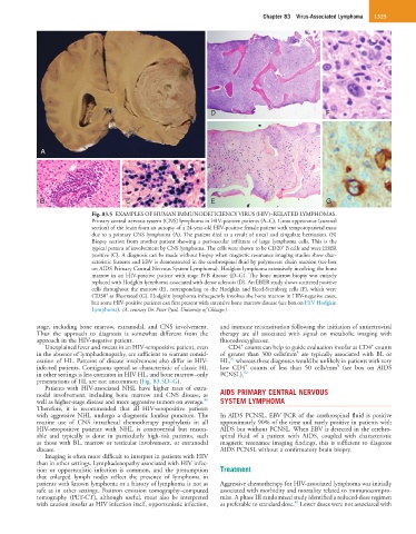
Fig. 83.5 EXAMPLES OF HUMAN IMMUNODEFICIENCY VIRUS (HIV)–RELATED LYMPHOMAS.
Primary central nervous system (CNS) lymphoma in HIV-positive patients (A–C). Gross appearance (coronal
section) of the brain from an autopsy of a 24-year-old HIV-positive female patient with temporoparietal mass
due to a primary CNS lymphoma (A). The patient died as a result of uncal and cingulate herniation. (B)
Biopsy section from another patient showing a perivascular infiltrate of large lymphoma cells. This is the
+
typical pattern of involvement by CNS lymphoma. The cells were shown to be CD20 B cells and were EBER
positive (C). A diagnosis can be made without biopsy when magnetic resonance imaging studies show char-
acteristic features and EBV is demonstrated in the cerebrospinal fluid by polymerase chain reaction (see box
on AIDS Primary Central Nervous System Lymphoma). Hodgkin lymphoma extensively involving the bone
marrow in an HIV-positive patient with stage IVB disease (D–G). The bone marrow biopsy was entirely
replaced with Hodgkin lymphoma associated with dense sclerosis (D). An EBER study shows scattered positive
cells throughout the marrow (E), corresponding to the Hodgkin and Reed-Sternberg cells (F), which were
+
CD30 as illustrated (G). Hodgkin lymphoma infrequently involves the bone marrow in HIV-negative cases,
but some HIV-positive patients can first present with extensive bone marrow disease (see box on HIV Hodgkin
Lymphoma). (A, courtesy Dr. Peter Pytel, University of Chicago.)
stage, including bone marrow, extranodal, and CNS involvement. and immune reconstitution following the initiation of antiretroviral
Thus the approach to diagnosis is somewhat different from the therapy are all associated with signal on metabolic imaging with
approach in the HIV-negative patient. fluorodeoxyglucose.
+
+
Unexplained fever and sweats in an HIV-seropositive patient, even CD4 counts can help to guide evaluation insofar as CD4 counts
3
in the absence of lymphadenopathy, are sufficient to warrant consid- of greater than 300 cells/mm are typically associated with BL or
81
eration of HL. Patterns of disease involvement also differ in HIV- HL, whereas these diagnoses would be unlikely in patients with very
+
3
infected patients. Contiguous spread so characteristic of classic HL low CD4 counts of less than 50 cells/mm (see box on AIDS
in other settings is less common in HIV HL, and bone marrow–only PCNSL). 82
presentations of HL are not uncommon (Fig. 83.5D–G).
Patients with HIV-associated NHL have higher rates of extra-
nodal involvement, including bone marrow and CNS disease, as AIDS PRIMARY CENTRAL NERVOUS
80
well as higher-stage disease and more aggressive tumors on average. SYSTEM LYMPHOMA
Therefore, it is recommended that all HIV-seropositive patients
with aggressive NHL undergo a diagnostic lumbar puncture. The In AIDS PCNSL, EBV PCR of the cerebrospinal fluid is positive
routine use of CNS intrathecal chemotherapy prophylaxis in all approximately 90% of the time and rarely positive in patients with
HIV-seropositive patients with NHL is controversial but reason- AIDS but without PCNSL. When EBV is detected in the cerebro-
able and typically is done in particularly high-risk patients, such spinal fluid of a patient with AIDS, coupled with characteristic
as those with BL, marrow or testicular involvement, or extranodal magnetic resonance imaging findings, this is sufficient to diagnose
disease. AIDS PCNSL without a confirmatory brain biopsy.
Imaging is often more difficult to interpret in patients with HIV
than in other settings. Lymphadenopathy associated with HIV infec-
tion or opportunistic infection is common, and the presumption Treatment
that enlarged lymph nodes reflect the presence of lymphoma in
patients with known lymphoma or a history of lymphoma is not as Aggressive chemotherapy for HIV-associated lymphoma was initially
safe as in other settings. Positron emission tomography–computed associated with morbidity and mortality related to immunocompro-
tomography (PET-CT), although useful, must also be interpreted mise. A phase III randomized study identified a reduced-dose regimen
83
with caution insofar as HIV infection itself, opportunistic infection, as preferable to standard dose. Lower doses were not associated with