
Page 1486 - Hematology_ Basic Principles and Practice ( PDFDrive )
P. 1486
Chapter 83 Virus-Associated Lymphoma 1321
A A C E E
B D FF
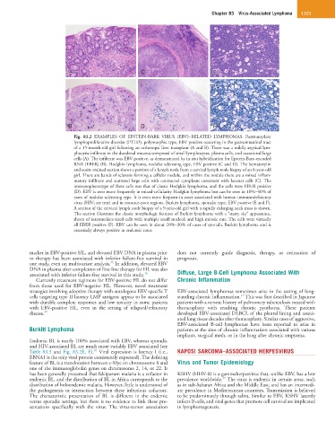
Fig. 83.2 EXAMPLES OF EPSTEIN-BARR VIRUS (EBV)–RELATED LYMPHOMAS. Posttransplant
lymphoproliferative disorder (PTLD), polymorphic type, EBV positive occurring in the gastrointestinal tract
of a 15-month-old girl following an orthotopic liver transplant (A and B). There was a mildly atypical lym-
phocytic infiltrate in the duodenal mucosa composed of small lymphocytes, plasma cells, and occasional large
cells (A). The infiltrate was EBV positive, as demonstrated by in situ hybridization for Epstein-Barr–encoded
RNA (EBER) (B). Hodgkin lymphoma, nodular sclerosing type, EBV positive (C and D). The hematoxylin
and eosin–stained section shows a portion of a lymph node from a cervical lymph node biopsy of an 8-year-old
girl. There are bands of sclerosis forming a cellular nodule, and within the nodule there are a mixed inflam-
matory infiltrate and scattered large cells with contracted cytoplasm consistent with lacunar cells (C). The
immunophenotype of these cells was that of classic Hodgkin lymphoma, and the cells were EBER positive
(D). EBV is seen more frequently in mixed-cellularity Hodgkin lymphoma but can be seen in 10%–40% of
cases of nodular sclerosing type. It is even more frequent in cases associated with human immunodeficiency
virus (HIV; see text) and in resource-poor regions. Burkitt lymphoma, sporadic type, EBV positive (E and F).
A section of the cervical lymph node biopsy of a 9-year-old girl with a rapidly enlarging neck mass is shown.
The section illustrates the classic morphologic features of Burkitt lymphoma with a “starry sky” appearance,
sheets of intermediate-sized cells with multiple small nucleoli and high mitotic rate. The cells were virtually
all EBER positive (F). EBV can be seen in about 20%–30% of cases of sporadic Burkitt lymphoma and is
essentially always positive in endemic cases.
marker in EBV-positive HL, and elevated EBV DNA in plasma prior does not currently guide diagnosis, therapy, or estimation of
to therapy has been associated with inferior failure-free survival in prognosis.
36
one study, even on multivariate analysis. In addition, elevated EBV
DNA in plasma after completion of first-line therapy for HL was also
associated with inferior failure-free survival in this study. 36 Diffuse, Large B-Cell Lymphoma Associated With
Currently treatment regimens for EBV-positive HL do not differ Chronic Inflammation
from those used for EBV-negative HL. However, novel treatment
strategies involving adoptive therapy with autologous EBV-specific T EBV-associated lymphomas sometimes arise in the setting of long-
15
cells targeting type II latency LMP antigens appear to be associated standing chronic inflammation. This was first described in Japanese
with durable complete responses and low toxicity in some patients patients with a remote history of pulmonary tuberculosis treated with
with EBV-positive HL, even in the setting of relapsed/refractory thoracoplasty with resulting chronic pyothorax. These patients
disease. 39 developed EBV-associated DLBCL of the pleural lining and associ-
ated lung tissue decades after thoracoplasty. Similar cases of aggressive,
EBV-associated B-cell lymphomas have been reported to arise in
Burkitt Lymphoma patients at the sites of chronic inflammation associated with various
implants, surgical mesh, or in the lung after chronic empyema.
Endemic BL is nearly 100% associated with EBV, whereas sporadic
and HIV-associated BL are much more variably EBV associated (see
40
Table 83.3 and Fig. 83.2E, F). Viral expression is latency I (i.e., KAPOSI SARCOMA–ASSOCIATED HERPESVIRUS
EBNA1 is the only viral protein consistently expressed). The defining
feature of BL is a translocation between c-Myc on chromosome 8 and Virus and Tumor Epidemiology
one of the immunoglobulin genes on chromosome 2, 14, or 22. It
has been generally presumed that falciparum malaria is a cofactor in KSHV (HHV-8) is a gammaherpesvirus that, unlike EBV, has a low
41
endemic BL, and the distribution of BL in Africa corresponds to the prevalence worldwide. The virus is endemic in certain areas, such
distribution of holoendemic malaria. However, little is understood of as in sub-Saharan Africa and the Middle East, and has an intermedi-
the pathogenesis or interaction between these infectious cofactors. ate prevalence in Mediterranean countries. Transmission is believed
The characteristic presentation of BL is different in the endemic to be predominately through saliva. Similar to EBV, KSHV latently
versus sporadic settings, but there is no evidence to link these pre- infects B cells, and viral genes that promote cell survival are implicated
sentations specifically with the virus. The virus-tumor association in lymphomagenesis.