
Page 1535 - Hematology_ Basic Principles and Practice ( PDFDrive )
P. 1535
1362 Part VII Hematologic Malignancies
may influence disease activity. Several reports suggested that exposure
to metals or their salts, pesticides or herbicides, and organic solvents
(halogenated or aromatic hydrocarbons) could be related to the
development of MF/SS. However, two well-designed case-control
studies have failed to support these observations.
Various theories have been advanced to explain the epidermotro-
pism of malignant T cells in MF/SS. Organ-specific affinity to skin
and other organs has been recognized in subsets of normal T cells.
Homing of CTCL cells to the skin is probably mediated by more
than one adhesion receptor mechanism. CTCL cells express cutane-
ous lymphocyte antigen, a skin-homing receptor that interacts with
E-selectin expressed by dermal venules. Furthermore, the cutaneous
lymphocyte antigens T lymphocytes also typically express the CCR4
chemokine receptor, which binds to chemokines produced by the
skin, such as the CC-chemokine ligands 17 and 22. Peripheral blood
mononuclear cells bind to cultured keratinocytes exposed to IFN-γ.
The major histocompatibility complex class II proteins along with
intercellular adhesion molecule 1 present on keratinocytes, and
attract and bind lymphocytes. Additional chemokine receptors, such
as CXC chemokine receptors 3 and 4, as well as unique integrins,
have been shown to have corresponding ligands or integrin receptors
on dermal Langerhans cells, suggesting a relationship between the
malignant T cells and host immune cells. The chemokine receptor
CCR4 is expressed by a spectrum of CTCL cells, and an anti-CCR4
monoclonal antibody has significant activity against these diseases.
An additional feature of MF/SS cells is the production of a
cytokine profile consistent with Th2 cells. Th2 cells produce IL-4,
IL-5, and IL-6, and they are inhibited by IFN-γ. The expression of
immunomodulatory molecule IL-17 is also increased in MF and SS.
Th2 cells are critical for stimulating antibody- and eosinophil- Fig. 85.10 ERYTHEMATOUS AND SCALY PATCH LESION OF
mediated responses. Hypergammaglobulinemia and eosinophilia are MYCOSIS FUNGOIDES.
occasionally seen in advanced cases of MF/SS and are consistent with
a Th2 profile. Stimulation of Th2 cells inhibits the Th1 subpopula-
tion of lymphocytes involved in cell-mediated immunity. Progression
of MF/SS is associated with immune suppression as a result of deple-
tion of this T-cell subset. The Th2 cytokine profile may explain the
decrease in tumor-infiltrating lymphocytes during tumor progression.
In addition to the effect of cytokines secreted by the neoplastic cells,
+
malignant CD4 cells express antigens (e.g., Fas ligand) that may
+
directly mediate elimination of the CD8 -infiltrating lymphocytes by
+
induction of apoptosis. CD4 T cells from MF skin lesions have an
effector memory phenotype, whereas T cells from SS skin lesions
display central memory characteristics with the expression of the
lymph node chemoattractant CCR7, which is not expressed in MF.
Clinical Presentation
Alibert reported the first case of MF in 1806. His patient developed
a skin eruption that progressed into mushroom-like tumors, prompt-
ing the term mycosis fungoides. Later in the 19th century, Bazin
defined the three classic cutaneous phases (patch, plaque, and tumor
stage) of the disease. The recognition of the clinical triad of intensely
pruritic erythroderma, lymphadenopathy, and abnormal hypercon-
voluted cells in the peripheral blood led to the description of SS.
MF is the prototype of CTCL observed in over 50% of CTCL
cases. The initial course of patients with MF is usually indolent.
Most patients give a history of antecedent skin lesions, usually non-
specific erythematous patches that can mimic eczema or psoriasis.
In many cases, there is an orderly progression from limited patches
to more generalized patches, plaques, tumors, and nodal or visceral
involvement. However, some patients may present with extensive
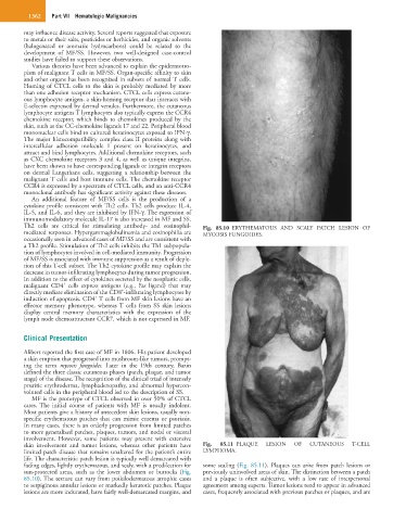
skin involvement and tumor lesions, whereas other patients have Fig. 85.11 PLAQUE LESION OF CUTANEOUS T-CELL
limited patch disease that remains unaltered for the patient’s entire LYMPHOMA.
life. The characteristic patch lesion is typically well demarcated with
fading edges, lightly erythematous, and scaly, with a predilection for some scaling (Fig. 85.11). Plaques can arise from patch lesions or
sun-protected areas, such as the lower abdomen or buttocks (Fig. previously uninvolved areas of skin. The distinction between a patch
85.10). The texture can vary from poikilodermatous atrophic cases and a plaque is often subjective, with a low rate of interpersonal
to serpiginous annular lesions or markedly keratotic patches. Plaque agreement among experts. Tumor lesions tend to appear in advanced
lesions are more indurated, have fairly well-demarcated margins, and cases, frequently associated with previous patches or plaques, and are