
Page 1536 - Hematology_ Basic Principles and Practice ( PDFDrive )
P. 1536
Chapter 85 T-Cell Lymphomas 1363
commonly associated with histologic evidence and large-cell transfor- The term large-plaque parapsoriasis has also been traditionally used
mation. They can be located on any part of the body. Ulceration of for these types of poorly defined lesions that may evolve into MF. It
these lesions is common, and secondary infection is a major cause most commonly consists of a few scattered, erythematous-to-brown
of morbidity (Fig. 85.12). Tumors may be the initial presentation plaques that are usually larger than 6 cm. There is a predilection for
in a small percentage of patients (d’emblée presentation, Vidal and the buttocks and intertriginous areas. Histologic examination shows
Brocq, 1889). a superficial lymphocytic infiltrate with minimal nuclear atypia.
SS patients present with generalized desquamative erythroderma, Epidermotropism is scant or absent, and dermal fibrosis correlates
pruritus, and circulating malignant cells. Peripheral blood usually with the chronicity of the process. Plaques can persist for decades
shows a significant number or percentage of hyperconvoluted atypical before a frank evolution to MF occurs. Approximately 10%–30% of
lymphocytes (Fig. 85.13). Approximately 5%–10% of all newly patients ultimately develop an overt malignant transformation.
reported cases of CTCL are SS. In its most advanced form, patients Large-plaque parapsoriasis is more likely to evolve into MF than
with SS suffer from alopecia, ectropion, leonine facies, hyperkeratosis, small-plaque lesions.
nail dystrophy, fissuring of the palms and soles, and severe pruritus
and cutaneous pain. Many other entities can clinically mimic this
disease, including drug eruptions, atopic dermatitis, contact derma- Idiopathic Follicular Mucinosis
titis, and erythrodermic psoriasis. A number of variant presentations
of CTCL are described in the following sections. Follicular mucinosis is also considered a variant of cutaneous lym-
phoid dyscrasias that manifests with grouped erythematous follicular
papules or boggy or indurated nodular plaques, notably devoid of
Cutaneous Lymphoid Dyscrasias (Clonal Dermatitis) hair (Fig. 85.14). There is a predilection for the head and neck area,
especially the forehead, which has the highest density of pilosebaceous
Cutaneous lymphoid dyscrasias or clonal dermatitis, which include a units. Histopathologic evaluation reveals cells in sebaceous glands
variety of lymphocyte-rich dermatoses, are often characterized by often associated with destruction of hair follicle structures due to
clonal T-lymphocyte proliferations that occasionally progress to bona infiltration by a T-lymphocytic process. This idiopathic condition can
fide mycosis fungoides. Clinically they exhibit a myriad of cutaneous evolve or be associated with folliculotropic MF. Even idiopathic cases
presentations from poikiloderma (atrophic patches) to hyperpig-
mented areas resembling pigmented purpuric dermatosis or an
acneiform presentation of follicular mucinosis.
Fig. 85.12 ULCERATED TUMORS ARISING FROM MYCOSIS FUN- Fig. 85.14 FOLLICULAR MUCINOSIS SHOWING A PATCH OF
GOIDES PLAQUES. ALOPECIA WITH FOLLICULAR PROMINENCE.
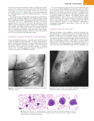
A A B C D
Fig. 85.13 SÉZARY CELLS. Peripheral smear (A) with Sézary cells associated with eosinophilia. Note the
cerebriform nuclei with fine chromatin (B–D). The hyperconvoluted nature of the nuclei is evident as complex
nuclear folds seen through the chromatin.