
Page 1613 - Hematology_ Basic Principles and Practice ( PDFDrive )
P. 1613
1434 Part VII Hematologic Malignancies
fraction. The patient was placed on lisinopril and did not improve.
He returned and underwent catheterization with coronary angiogra-
phy. His coronary arteries were normal, and he was diagnosed with
deconditioning. His dyspnea continued, and he was referred to a
pulmonologist, who recognized the long-standing monoclonal gam-
mopathy and obtained light chain studies that showed a λ light chain
of 41 mg/dL and κ of 10 mg/dL. The patient underwent a subcuta-
neous fat aspiration that demonstrated amyloid deposits, which, by
mass spectroscopy, were found to be of λ origin. At this point, the
patient had New York Heart Association class IV heart failure; he
died within 3 months thereafter.
Comment on Patient 2
This case is an example of a classic failure to recognize that patients
with monoclonal gammopathies should have amyloidosis included in
the differential diagnosis. The fact that the monoclonal protein was
stable would certainly argue against the development of multiple
myeloma, but light-chain amyloidosis regularly occurs without any
change in the M component over time. The failure to recognize the
patient’s progressive fatigue and shortness of breath as cardiac amyloid
is not unusual. The echocardiographic findings of thickening were
interpreted as being caused by hypertension, but, in fact, they were
caused by infiltrative cardiomyopathy, and the cardiac catheterization
and angiogram without an endomyocardial biopsy led to a missed
diagnosis. Only an increased index of suspicion in patients with a
known monoclonal protein allowed this diagnosis to be confirmed.
One of the major difficulties in the diagnosis of amyloidosis is
that there is virtually no blood test or imaging study that is diagnostic
of the disease. The most common symptoms associated with amyloi-
dosis are fatigue, lower extremity edema (may be cardiac or renal),
unexplained weight loss (often leading to search for occult malig-
nancy), exertional dyspnea, orthostatic hypotension, and paresthesias.
These symptoms are quite vague, and in a general medical practice,
there are hundreds of disorders that are far more common and are
responsible for light-chain amyloidosis. As noticed in Patient 2, the
fatigue, which is often caused by early cardiac amyloid but is not
associated with overt congestive heart failure, can be missed. Cardiac Fig. 88.3 ENLARGED TONGUE INFILTRATED BY AMYLOID
amyloidosis is a classic form of heart failure with preserved ejection DEPOSITS.
fraction owing to the restrictive physiology associated with
amyloidosis.
Edema in amyloidosis may be a manifestation of high right-sided
filling pressures, leading to lower extremity edema, or it may be a
consequence of renal involvement with the nephrotic syndrome
(peripheral edema, hypoalbuminemia, hyperlipidemia, and protein-
uria). Even when nephrotic range proteinuria is seen, amyloidosis,
which is known to cause nephrotic syndrome in 10% of adults who
are nondiabetic, is infrequently considered in the differential diagno-
sis, which would include minimal change glomerulopathy, as well as
membranous and membranoproliferative glomerulopathy.
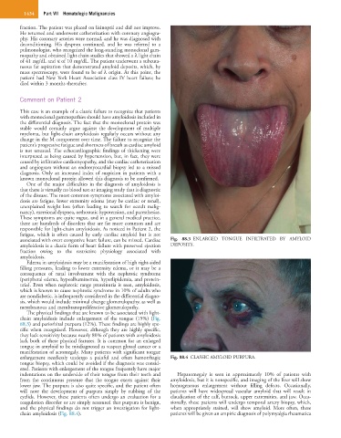
The physical findings that are known to be associated with light-
chain amyloidosis include enlargement of the tongue (15%) (Fig.
88.3) and periorbital purpura (12%). These findings are highly spe-
cific when recognized. However, although they are highly specific,
they lack sensitivity because nearly 80% of patients with amyloidosis
lack both of these physical features. It is common for an enlarged
tongue in amyloid to be misdiagnosed as suspect glossal cancer or a
manifestation of acromegaly. Many patients with significant tongue
enlargement needlessly undergo a painful and often hemorrhagic Fig. 88.4 CLASSIC AMYLOID PURPURA.
tongue biopsy, which could be avoided if the diagnosis was consid-
ered. Patients with enlargement of the tongue frequently have major
indentations on the underside of their tongue from their teeth and Hepatomegaly is seen in approximately 10% of patients with
from the continuous pressure that the tongue exerts against their amyloidosis, but it is nonspecific, and imaging of the liver will show
lower jaw. The purpura is also quite specific, and the patient often homogeneous enlargement without filling defects. Occasionally,
will note the development of purpura simply by rubbing of the patients will have widespread vascular amyloid that will result in
eyelids. However, these patients often undergo an evaluation for a claudication of the calf, buttock, upper extremities, and jaw. Occa-
coagulation disorder or are simply reassured that purpura is benign, sionally, these patients will undergo temporal artery biopsy, which,
and the physical findings do not trigger an investigation for light- when appropriately stained, will show amyloid. More often, these
chain amyloidosis (Fig. 88.4). patients will be given an empiric diagnosis of polymyalgia rheumatica