
Page 1616 - Hematology_ Basic Principles and Practice ( PDFDrive )
P. 1616
Chapter 88 Immunoglobulin Light Chain Amyloidosis (Primary Amyloidosis) 1437
The typical sites associated with localized amyloidosis include skin,
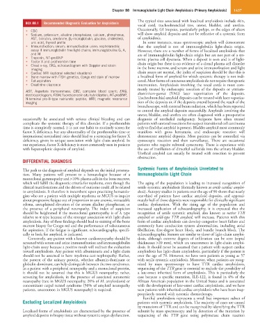
BOX 88.1 Recommended Diagnostic Evaluation for Amyloidosis
vocal cord, tracheobronchial tree, ureter, bladder, and urethra.
• CBC Occasionally, GI biopsies, particularly polyps, or the edges of ulcers
• Sodium, potassium, alkaline phosphatase, calcium, phosphorus, will show amyloid deposits and not be reflective of a systemic form
AST, bilirubin, creatinine, β 2 -microglobulin, glucose, cholesterol, of amyloidosis.
uric acid, thyroid profile In some instances, mass spectroscopic analysis will demonstrate
• Immunofixation; serum; immunofixation urine; nephelometric that the amyloid is not of immunoglobulin light-chain origin.
assay if immunoglobulin free light chains; immunoglobulins G, A, However, there are a number of forms of localized amyloidosis that
and M are of immunoglobulin light-chain origin but are not part of a sys-
• Troponin, NT-proBNP temic plasma cell dyscrasia. When a deposit is seen and is of light-
• Factor X and prothrombin time chain origin but there is no evidence of a clonal plasma cell disorder
• Chest x-ray, EKG, echocardiogram with Doppler and strain
imaging in the bone marrow, and serum and urine immunofixation and light
• Cardiac MRI (optional selected situations) chain assays are normal, the index of suspicion should be that this is
• Bone marrow with FISH genetics, Congo red stain of marrow a localized form of amyloid for which systemic therapy is not indi-
• Fat aspiration cated. Most forms of cutaneous amyloidosis do not require therapeutic
• Creatinine clearance intervention. Amyloidosis involving the vocal cords is most com-
monly treated by endoscopic resection of the deposits or yttrium-
AST, Aspartate transaminase; CBC, complete blood count; EKG,
electrocardiogram; FISH, fluorescence in situ hybridization; NT-proBNP, aluminium-garnet (YAG) laser vaporization of the deposits.
N-terminal pro-B-type natriuretic peptide; MRI, magnetic resonance Tracheobronchial amyloid deposits can be treated with laser vaporiza-
imaging. tion of the deposits or, if the deposits extend beyond the reach of the
bronchoscope, with external beam radiation, which has been reported
to control the amyloid deposits successfully. Amyloids involving the
ureter, bladder, and urethra are often diagnosed with a preoperative
occasionally be associated with serious clinical bleeding and can diagnosis of urothelial malignancy. Surgeons have often treated
complicate the systemic therapy of this disorder. If a prothrombin patients with ureteral resections for suspect transitional cell carcinoma
time is completely normal, it is not our habit to routinely screen for only to find that amyloid is present. Bladder amyloid most commonly
factor X deficiency, but any abnormality of the prothrombin time or manifests with gross hematuria, and endoscopic resection will
international normalized ratio should lead to a screening for factor X demonstrate amyloid deposits. Most patients can be controlled via
deficiency, given its unique association with light chain amyloid. In cystoscopic resection and then surveillance. There are occasional
our experience, factor X deficiency is most commonly seen in patients patients who require subtotal cystectomy. There is experience with
with hepatosplenic deposits of amyloid. the use of instillation of dimethyl sulfoxide into the urinary bladder.
Urethral amyloid can usually be treated with resection to prevent
obstruction.
DIFFERENTIAL DIAGNOSIS
Systemic Forms of Amyloidosis Unrelated to
The path to the diagnosis of amyloid depends on the initial presenta-
tion. Many patients will present to a hematologist because of a Immunoglobulin Light Chain
monoclonal gammopathy and >10% plasma cells in the bone marrow,
which will fulfill the diagnostic criteria for myeloma, even though the The aging of the population is leading to increased recognition of
clinical manifestations and the drivers of outcome could all be related senile systemic amyloidosis (formally known as senile cardiac amyloi-
to amyloidosis. It therefore is incumbent upon practicing hematolo- dosis). Autopsy studies in patients over the age of 90 shows that nearly
gists who see a patient with multiple myeloma or MGUS to inquire one-third of patients have cardiac amyloid deposits, and approxi-
about progressive fatigue out of proportion to any anemia, intractable mately half of those deposits were responsible for clinically significant
edema, unexplained elevation of the serum alkaline phosphatase, or cardiac dysfunction. With the rising age of the population and
the presence of a peripheral neuropathy. The index of suspicion increased application of echocardiography, it is expected that the
should be heightened if the monoclonal gammopathy is of λ type recognition of senile systemic amyloid, also known as native TTR
relative to κ type because of the stronger association with light chain amyloid or wild-type TTR amyloid, will increase. Patients with this
amyloidosis. Any of these findings should lead to staining of the bone form of cardiac amyloidosis can develop congestive heart failure and
marrow biopsy for Congo red and the performance of subcutaneous commonly have conduction system abnormalities, including atrial
fat aspiration. If the fatigue is significant, echocardiography, specifi- fibrillation, first-degree heart block, and bundle branch block. The
cally to look for amyloid, is indicated. echocardiographic features are similar to those of light-chain amyloi-
Conversely, any patient with a known cardiomyopathy should be dosis, although extreme degrees of infiltration can be seen (septal
screened with serum and urine immunofixation and immunoglobulin thicknesses >20 mm), which are uncommon in light-chain amyloi-
light-chain assay because a positive result will redirect the evaluation dosis. It should never be assumed that a patient with suspect cardiac
toward amyloidosis. Any patient with albuminuria and a light chain amyloidosis has light-chain amyloidosis, particularly if the patient is
should not be assumed to have myeloma cast nephropathy. Rather, over the age of 70. However, we have seen patients as young as 57
the pattern of the urinary protein, whether albumin-dominant or with senile systemic amyloidosis. Moreover, when patients are recog-
globulin-dominant, can help distinguish between the two syndromes. nized by mass spectroscopy to have TTR cardiac amyloidosis,
In a patient with a peripheral neuropathy and a monoclonal protein, sequencing of the TTR gene is essential to exclude the possibility of
it should not be assumed that this is MGUS neuropathy; rather, a late-onset inherited form of amyloidosis. This is particularly the
screening for amyloidosis, in the presence of associated autonomic case because a specific mutation, ILE-122, is found in 4% of the
neuropathy (rare in MGUS neuropathy, 20% of AL amyloidosis) or African American population in the United States and is associated
concomitant carpal tunnel syndrome (50% of amyloid neuropathy with the development of late-onset cardiac amyloidosis, and we have
patients, uncommon in MGUS neuropathy) is required. seen patients with inherited cardiac amyloidosis who have been inap-
propriately treated with systemic chemotherapy.
Familial amyloidosis represents a small but important subset of
Excluding Localized Amyloidosis patients with systemic amyloidosis. The majority of cases are caused
by mutations of TTR and can be recognized by identifying the TTR
Localized forms of amyloidosis are characterized by the presence of subunit by mass spectroscopy and by detection of the mutation by
amyloid deposits in biopsy tissue without systemic organ dysfunction. sequencing of the TTR gene using polymerase chain reaction