
Page 1615 - Hematology_ Basic Principles and Practice ( PDFDrive )
P. 1615
1436 Part VII Hematologic Malignancies
TABLE Nomenclature of Amyloidosis
88.2
Protein Precursor Clinical Characteristics
AL or AH Immunoglobulin light Primary or localized; myeloma or
or heavy chain macroglobulinemia associated
AA SAA Secondary or familial
Mediterranean fever, familial
periodic fever syndromes
ATTR Transthyretin Familial and senile
A fibrinogen Fibrinogen Familial renal amyloidosis
(Ostertag type)
Aβ 2 M β 2 -Microglobulin Dialysis associated; carpal
tunnel syndrome
Aβ ABPP Alzheimer disease
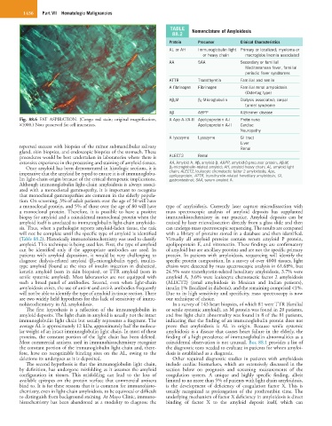
Fig. 88.6 FAT ASPIRATION. (Congo red stain; original magnification, A Apo A-I/A-II Apolipoprotein A-I Proteinuria
×1000.) Note preserved fat cell interstices. Apolipoprotein A-II Cardiac
Neuropathy
A lysozyme Lysozyme GI tract
Liver
reported success with biopsies of the minor submandibular salivary Renal
gland, skin biopsies, and endoscopic biopsies of the stomach. These
procedures would be best undertaken in laboratories where there is ALECT2 Renal
extensive experience in the processing and staining of amyloid tissues. AA, Amyloid A; Aβ, amyloid-β; ABPP, amyloid-β precursor protein; Aβ 2M,
Once amyloid has been demonstrated in histologic sections, it is β 2-microglobulin-related amyloid; AH, amyloid heavy chain; AL, amyloid light
imperative that the amyloid be typed to ensure it is of immunoglobu- chain; ALECT2, leukocyte chemotactic factor 2 amyloidosis; Apo,
apolipoprotein; ATTR, transthyretin-related hereditary amyloidosis; GI,
lin light-chain origin because of the critical therapeutic implications. gastrointestinal; SAA, serum amyloid A.
Although immunoglobulin light-chain amyloidosis is always associ-
ated with a monoclonal gammopathy, it is important to recognize
that monoclonal gammopathies are common in the elderly popula-
tion. On screening, 3% of adult patients over the age of 50 will have
a monoclonal protein, and 5% of those over the age of 80 will have type of amyloidosis. Currently laser capture microdissection with
a monoclonal protein. Therefore, it is possible to have a positive mass spectroscopic analysis of amyloid deposits has supplanted
biopsy for amyloid and a coincidental monoclonal protein when the immunohistochemistry in our practice. Amyloid deposits can be
amyloid itself is unrelated to immunoglobulin light-chain amyloido- excised by laser microdissection directly from a glass slide and then
sis. Thus, when a pathologist reports amyloid-laden tissue, the task can undergo mass spectroscopic sequencing. The results are compared
will not be complete until the specific type of amyloid is identified with a library of proteins stored in a database and then identified.
(Table 88.2). Historically immunohistochemistry was used to classify Virtually all amyloid proteins contain serum amyloid P protein,
amyloid. This technique is being used less. First, the type of amyloid apolipoprotein E, and vitronectin. These findings are confirmatory
can be identified only if the appropriate antibodies are used. In of amyloid but are ancillary proteins and are not the specific primary
patients with amyloid deposition, it would be very challenging to protein. In patients with amyloidosis, sequencing will identify the
diagnose dialysis-related amyloid (β 2 -microglobulin type), insulin- specific protein composition. In a survey of over 4000 tissues, light
type amyloid (found at the sites of insulin injection in diabetics), chains were detected by mass spectroscopic analysis in 61.68%, but
keratin amyloid (seen in skin biopsies), or TTR amyloid (seen in 24.5% were transthyretin-related hereditary amyloidosis, 3.7% were
senile systemic amyloid). Most laboratories are not equipped with amyloid A, 3.6% were leukocyte chemotactic factor 2 amyloidosis
such a broad panel of antibodies. Second, even when light-chain (ALECT2) (renal amyloidosis in Mexican and Indian patients),
amyloidosis exists, the use of anti-κ and anti-λ antibodies frequently insulin 1% (localized in diabetic), and the remaining comprised <1%.
will not be able to identify the type of amyloid in tissue section. There Due to its high sensitivity and specificity, mass spectroscopy is now
are two widely held hypotheses for this lack of sensitivity of immu- our technique of choice.
nohistochemistry in AL amyloidosis. In a survey of 143 heart biopsies, of which 81 were TTR (familial
The first hypothesis is a reflection of the immunoglobulin in or senile systemic amyloid), an M protein was found in 20 patients,
amyloid deposits. The light chain in amyloid is usually not the intact and free light chain abnormality was found in 8 of the 81 patients,
immunoglobulin light chain but usually represents a fragment. The indicating that the finding of an immunoglobulin protein does not
average AL is approximately 12 kDa, approximately half the molecu- prove that amyloidosis is AL in origin. Because senile systemic
lar weight of an intact immunoglobulin light chain. In most of these amyloidosis is a disease that causes heart failure in the elderly, the
proteins, the constant portion of the light chain has been deleted. finding of a high prevalence of immunoglobulin abnormalities as a
Most commercial antisera used in immunohistochemistry recognize coincidental observation is not unusual. Box 88.1 provides a list of
the constant portion of the immunoglobulin light chain and, there- the diagnostic tests needed to evaluate in patients for whom amyloi-
fore, have no recognizable binding sites on the AL, owing to the dosis is established as a diagnosis.
deletions its undergoes as it is deposited. Other required diagnostic studies in patients with amyloidosis
The second hypothesis is that the immunoglobulin light chain, include cardiac biomarkers, which are extensively discussed in the
by definition, has undergone misfolding as it assumes the amyloid section below on prognosis and screening measurements of the
configuration in tissues. This misfolding can lead to the loss of coagulation system. A unique and highly specific finding, albeit
available epitopes on the protein surface that commercial antisera limited to no more than 5% of patients with light chain amyloidosis,
bind to. It is for these reasons that it is common for immunohisto- is the development of deficiency of coagulation factor X. This is
chemistry, even in light-chain amyloidosis, to be equivocal or difficult usually recognized as prolongation of the prothrombin time. The
to distinguish from background staining. At Mayo Clinic, immuno- underlying mechanism of factor X deficiency in amyloidosis is direct
histochemistry has been abandoned as a modality to diagnose the binding of factor X to the amyloid deposit itself, which can