
Page 1735 - Hematology_ Basic Principles and Practice ( PDFDrive )
P. 1735
1540 Part IX Cell-Based Therapies
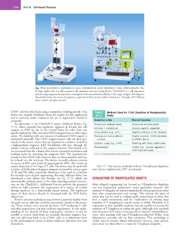
Fig. 97.2 MILTENYI CLINIMACS CELL SEPARATOR AND PRODIGY CELL PROCESSOR. The
Prodigy (right side) is a self-contained cell separation and processing device. CliniMACS is a cell separation
system using magnetic nanoparticles conjugated with monoclonal antibody to the target antigen. Investigators
should determine the status of regulatory approval of these devices before clinical use. (Copyright 2015 Miltenyi
Biotec GmbH. All rights reserved.)
+
CD34 cells from the beads using a competitive binding peptide. This TABLE Methods Used for T-Cell Depletion of Hematopoietic
device was recently withdrawn from the market for this application 97.2 Grafts
and is currently under evaluation for use in regenerative medicine
protocols. Destruction in Situ Physical Separation
An alternative is the CliniMACS system (Miltenyi Biotec; Fig. Monoclonal antibody-based Monoclonal antibody-based
97.2), which currently has regulatory approval in Europe but still Antibody + complement Immunomagnetic separation
requires an IND for use in the United States for other than one
specific indication. This uses anti-CD34 nanoparticles to effect sepa- Immunotoxins (e.g., ricin) Negative selection (T-cell removal)
ration. The labeling with and removal of unbound CD34 reagent is Panning and immunoaffinity Positive selection (CD34 selection)
performed manually. The CD34 reagent-treated cells are then pro- columns CliniMACs device
cessed on the device, where they are retained on a column located in Cytotoxic drugs (e.g., 4-HC) Rosetting with sheep erythrocytes
a high-gradient magnetic field. Nonlabeled cells flow through the
column and are collected in the negative fraction. The labeled cells Photopheresis Lectins (e.g., soybean agglutinin)
are recovered from the column after several automated separation and Centrifugal elutriation
washing cycles by removing the magnetic field. The nanoparticles 4-HC, 4-Hydroperoxycyclophosphamide.
+
remain on the CD34 cells; however, they are biocompatible and may
be infused into the recipient. The device normally achieves purities
in excess of 90% with yields of approximately 60%. This results in
passive depletion of 4–6 logs of T cells. The device may be used with Table 97.2 lists various methods of direct T-lymphocyte depletion
a variety of MAbs directed against antigens expressed by various types and indirect depletion by HPC enrichment.
(T, B, and NK cells), potentially allowing it to be used as a platform
for multiple types of graft engineering. Recently, Miltenyi Biotec has
introduced a new device (the Prodigy; see Fig. 97.2), which can EVALUATION OF MANIPULATED GRAFTS
automatically perform many of the steps requiring manual interven-
tion on the CliniMACs. Additional features provide the potential Most allograft engineering has focused on T-lymphocyte depletion
ability to fully automate the preparation of a variety of cellular and has emphasized quantitative versus qualitative removal. The
therapy products in a functionally closed system. The regulatory majority of allografts are infused immediately after preparation rather
status of these devices should be discussed with the FDA before than after cryopreservation and storage. This restricts the types of
clinical use. assays that can be used to evaluate graft composition to those that
Positive selection techniques may, however, passively deplete from have a rapid turnaround, and the implications of infusing large
the graft certain cells that could be of potential benefit to the recipi- numbers of T lymphocytes can be severe or lethal. Therefore, it is
ent. These include some stromal elements, GVT-mediating T cells, important to have available methods that can rapidly enumerate the
and other populations that may facilitate engraftment. As our under- numbers of T lymphocytes within the graft. Although early methods
standing of the identity of these populations improves, it may be used detection of E-rosette–forming cells or manual immunofluores-
possible to recover them from the normally discarded negative frac- cence after staining with pan–T-lymphocyte–directed MAbs, most
+
tion and add them back to the CD34 cells, or to administer them laboratories currently rely on flow cytometry. This technology is
in the posttransplant period as donor leukocyte infusions (see later widely used in routine clinical laboratories; however, some precau-
discussion). tions must be taken when it is used for T-depleted allografts.