
Page 1737 - Hematology_ Basic Principles and Practice ( PDFDrive )
P. 1737
1542 Part IX Cell-Based Therapies
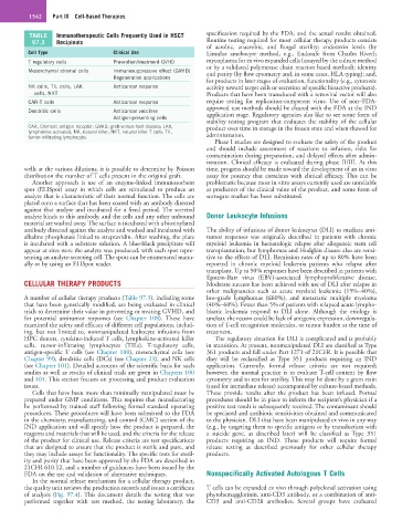
TABLE Immunotherapeutic Cells Frequently Used in HSCT specification required by the FDA, and the actual results obtained.
97.3 Recipients Routine testing required for most cellular therapy products consists
of aerobic, anaerobic, and fungal sterility; endotoxin levels (by
Cell Type Clinical Use Limulus amebocyte method, e.g., Endosafe from Charles River);
T regulatory cells Prevention/treatment GVHD mycoplasma for ex vivo expanded cells (assayed by the culture method
or by a validated polymerase chain reaction based method); identity
Mesenchymal stromal cells Immunosuppressive effect (GVHD) and purity (by flow cytometry and, in some cases, HLA typing); and,
Regenerative applications
for products in later stages of evaluation, functionality (e.g., cytotoxic
NK cells, TIL cells, LAK Anticancer response activity toward target cells or secretion of specific bioactive products).
cells, NKT Products that have been transduced with a retroviral vector will also
CAR-T cells Anticancer response require testing for replication-competent virus. Use of non–FDA-
approved test methods should be cleared with the FDA at the IND
Dendritic cells Anticancer vaccines application stage. Regulatory agencies also like to see some form of
Antigen-presenting cells
stability testing program that evaluates the stability of the cellular
CAR, Chimeric antigen receptor; GVHD, graft-versus-host disease; LAK, product over time in storage in the frozen state and when thawed for
lymphokine activated; NK, natural killer; NKT, natural killer T cells; TIL, administration.
tumor-infiltrating lymphocyte.
Phase I studies are designed to evaluate the safety of the product
and should include assessment of reactions to infusion, risks for
contamination during preparation, and delayed effects after admin-
istration. Clinical efficacy is evaluated during phase II/III. At this
wells at the various dilutions, it is possible to determine by Poisson time, progress should be made toward the development of an in vitro
distribution the number of T cells present in the original graft. assay for potency that correlates with clinical efficacy. This can be
Another approach is use of an enzyme-linked immunosorbent problematic because most in vitro assays currently used are unreliable
spot (ELISpot) assay in which cells are stimulated to produce an as predictors of the clinical value of the product, and some form of
analyte that is characteristic of their normal function. The cells are surrogate marker has been substituted.
plated onto a surface that has been coated with an antibody directed
against that analyte and incubated for a fixed period. The secreted
analyte binds to this antibody, and the cells and any other unbound Donor Leukocyte Infusions
material are washed away. The surface is incubated with a biotinylated
antibody directed against the analyte and washed and incubated with The ability of infusions of donor leukocytes (DLI) to mediate anti-
alkaline phosphatase linked to streptavidin. After washing, the plate tumor responses was originally described in patients with chronic
is incubated with a substrate solution. A blue-black precipitate will myeloid leukemia in hematologic relapse after allogeneic stem cell
appear at sites were the analyte was produced, with each spot repre- transplantation, but lymphomas and Hodgkin disease also are sensi-
senting an analyte-secreting cell. The spots can be enumerated manu- tive to the effects of DLI. Remission rates of up to 80% have been
ally or by using an ELISpot reader. reported in chronic myeloid leukemia patients who relapse after
transplant. Up to 90% responses have been described in patients with
Epstein-Barr virus (EBV)-associated lymphoproliferative disease.
CELLULAR THERAPY PRODUCTS Moderate success has been achieved with use of DLI after relapse in
other malignancies such as acute myeloid leukemia (15%–40%),
A number of cellular therapy products (Table 97.3), including some low-grade lymphomas (≤60%), and metastatic multiple myeloma
that have been genetically modified, are being evaluated in clinical (40%–60%). Fewer than 5% of patients with relapsed acute lympho-
trials to determine their value in preventing or treating GVHD, and blastic leukemia respond to DLI alone. Although the etiology is
for potential antitumor responses (see Chapter 108). These have unclear, the reason could be lack of antigenic expression, downregula-
examined the safety and efficacy of different cell populations, includ- tion of T-cell recognition molecules, or tumor burden at the time of
ing, but not limited to, nonmanipulated leukocyte infusions from treatment.
HPC donors, cytokine-induced T cells, lymphokine-activated killer The regulatory situation for DLI is complicated and is probably
cells, tumor-infiltrating lymphocytes (TILs), T-regulatory cells, in transition. At present, nonmanipulated DLI are classified as Type
antigen-specific T cells (see Chapter 100), mesenchymal cells (see 361 products and fall under Part 1271 of 21CFR. It is possible that
Chapter 99), dendritic cells (DCs) (see Chapter 23), and NK cells they will be reclassified as Type 351 products requiring an IND
(see Chapter 101). Detailed accounts of the scientific basis for such application. Currently, formal release criteria are not required;
studies as well as results of clinical trials are given in Chapters 100 however, the normal practice is to evaluate T-cell content by flow
and 101. This section focuses on processing and product evaluation cytometry and to test for sterility. This may be done by a gram stain
issues. (used for immediate release) accompanied by culture-based methods.
Cells that have been more than minimally manipulated must be These provide results after the product has been infused. Formal
prepared under GMP conditions. This requires that manufacturing procedures should be in place to inform the recipient’s physician if a
be performed by trained staff following formal standard operating positive test result is subsequently received. The contaminant should
procedures. These procedures will have been submitted to the FDA be speciated and antibiotic sensitivities obtained and communicated
in the chemistry, manufacturing, and control (CMC) section of the to the physician. DLI that have been manipulated ex vivo in any way
IND application and will specify how the product is prepared, the (e.g., by targeting them to specific antigens or by transduction with
reagents and materials that will be used, and the criteria for the release a suicide gene, as described later) will be classified as Type 351
of the product for clinical use. Release criteria are test specifications products requiring an IND. These products will require formal
that are designed to ensure that the product is sterile and pure, and release testing as described previously for other cellular therapy
they may include assays for functionality. The specific tests for steril- products.
ity and purity that have been approved by the FDA are described in
21CFR 610.12, and a number of guidances have been issued by the
FDA on the use and validation of alternative techniques. Nonspecifically Activated Autologous T Cells
In the normal release mechanism for a cellular therapy product,
the quality unit reviews the production records and issues a certificate T cells can be expanded ex vivo through polyclonal activation using
of analysis (Fig. 97.4). This document details the testing that was phytohemagglutinin, anti-CD3 antibody, or a combination of anti-
performed together with test method, the testing laboratory, the CD3 and anti-CD28 antibodies. Several groups have evaluated