
Page 1895 - Hematology_ Basic Principles and Practice ( PDFDrive )
P. 1895
Chapter 109 Complications After Hematopoietic Cell Transplantation 1677
clinical criteria that include presence of jaundice, plus either hepato- polyribonucleotide with fibrinolytic, antithrombotic, and antiisch-
megaly, weight gain, and/or ascites within 2–3 weeks of stem cell emic properties, has been favorable with 30% to 40% of patients with
infusion. However, other causes of hyperbilirubinemia and weight severe SOS achieving complete resolution and improvement in sur-
26
gain early after transplantation (e.g., drugs, hepatitis, capillary leak, vival. Its efficacy in the prophylaxis and management of severe SOS
cardiac failure, volume overload) can complicate the differential is currently being investigated in larger clinical trials.
diagnosis, particularly for milder or less abrupt presentations of these
symptoms. Percutaneous or transabdominal needle biopsy of the liver
is hazardous in severely thrombocytopenic transplant recipients and Interstitial Pneumonitis
should be avoided. Transvenous biopsies may provide sufficient his-
tologic material for diagnosis and may allow determination of hepatic Interstitial pneumonitis is a common and frequently fatal complica-
wedge pressure product (greater than 10 mm is associated with SOS), tion, affecting up to 35% of allogeneic transplant recipients, although
but may be associated with hemorrhagic complications as well. recent advances in supportive care have substantially reduced this risk.
Ultrasonographic Doppler flow studies demonstrating reversal of Interstitial pneumonitis is notably less common after autografting. It
portal flow or a higher portal vein resistive index have been suggested is characterized by diffuse, interstitial inflammation accompanied by
as a noninvasive means of confirming the diagnosis, but their validity hypoxemia, dyspnea, and nonproductive cough, sometimes with fever.
has recently been questioned. SOS can be graded from mild to severe Risk factors associated with the development of interstitial pneumoni-
depending on the degree of hyperbilirubinemia and weight gain. tis include use of methotrexate for GVHD prophylaxis, older age at
Severe SOS is almost universally fatal within several weeks of onset. transplant, severe GVHD, interval from diagnosis of hematologic
Effective methods for prevention and treatment of SOS have not disease to HCT of 6 months or greater, poor pretransplant performance
been defined. Limited understanding of the cellular and microvascu- status and use of higher TBI dose rate (>4 cGy/min). Remarkably, in
lar pathophysiology of SOS has confounded development of more one study, the reported risk of interstitial pneumonitis was 8% when
rational approaches to its prevention and treatment. Possible none of these risk factors were present, compared with 94% when all
approaches include preventive therapy with low-dose heparin, pros- six factors were present. It has been hypothesized that URD transplan-
taglandin E, pentoxifylline (a TNF-α blocking agent), although none tation is more immunosuppressive and thus associated with more
has proved effective in carefully performed prospective trials. Post- severe opportunistic infections and greater risk of interstitial pneumo-
HCT hepatic injury including SOS has become less frequent with nitis, but this has not been rigorously investigated.
widespread and prolonged administration of ursodiol as a choleretic The course of interstitial pneumonitis is often catastrophic, mani-
and hepatic protectant. Recombinant tissue plasminogen factor has festing with rapidly progressive tachypnea, hypoxemia, and hemody-
been used successfully to treat established SOS, however, thrombolyt- namic compromise. Therefore, therapeutic intervention most
ics are associated with substantially increased risk of hemorrhage. frequently occurs before the return of definitive results of diagnostic
Transjugular intrahepatic portosystemic shunts have also been used tests and must be initiated based on the assessment of clinical risk
with some success. Early experience with defibrotide, a single-stranded factors and the underlying clinical setting (Box 109.3).
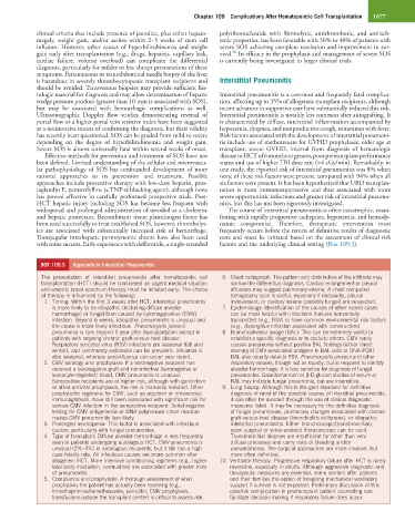
BOX 109.3 Approach to Interstitial Pneumonitis
The presentation of interstitial pneumonitis after hematopoietic cell 6. Chest radiograph: The pattern and distribution of the infiltrate may
transplantation (HCT) should be considered an urgent medical situation narrow the differential diagnosis. Cardiac enlargement or pleural
and empiric broad-spectrum therapy must be initiated early. The choice effusions may suggest pulmonary edema. A chest computed
of therapy is influenced by the following: tomography scan is useful, especially if nodularity, pleural
1. Timing: Within the first 3 weeks after HCT, interstitial pneumonitis involvement, or cavitary lesions (possibly fungal) are suspected.
is more likely to be idiopathic (including diffuse alveolar 7. Epidemiology: Identification of the causes of other recent cases
hemorrhage) or fungal than caused by cytomegalovirus (CMV) can be most helpful with infections that are horizontally
infection. Beyond 6 weeks, idiopathic pneumonitis is unusual and transmitted (e.g., RSV) or have common environmental risk factors
the cause is more likely infectious. Pneumocystis jirovecii (e.g., Aspergillus infection associated with construction).
pneumonia is rare beyond 1 year after transplantation except in 8. Bronchoalveolar lavage (BAL): This can be extremely useful to
patients with ongoing chronic graft-versus-host disease. establish a specific diagnosis or to exclude others. CMV rarely
Respiratory syncytial virus (RSV) infections are seasonal (fall and causes pneumonia without positive BAL findings (either direct
winter), and community outbreaks can be prevalent. Influenza is staining of CMV-associated antigens in BAL cells or DNA-PCR).
also seasonal, whereas parainfluenza can occur year round. BAL also usually detects RSV, Pneumocystis jirovecii and other
2. CMV serology and prophylaxis: If a seronegative recipient has respiratory viruses, though not as rapidly, but is required to identify
received a seronegative graft and noninfective (seronegative or alveolar hemorrhage. It is less sensitive for diagnosis of fungal
leukocyte-depleted) blood, CMV pneumonia is unusual. pneumonias. Galactomannan or β-D-glucan studies of serum or
Seropositive recipients are at higher risk, although with ganciclovir BAL may indicate fungal pneumonia, but are insensitive.
or other antiviral prophylaxis, the risk is markedly reduced. Other 9. Lung biopsy: Although this is the gold standard for definitive
prophylactic regimens for CMV, such as acyclovir or intravenous diagnosis of most of the possible causes of interstitial pneumonitis,
immunoglobulin, have still been associated with significant risk for it can often be avoided through the use of clinical diagnostic
serious CMV infection in the seropositive recipient. Serial negative measures listed. It may be necessary for the definitive diagnosis
testing for CMV antigenemia or DNA-polymerase chain reaction of fungal pneumonias, pulmonary changes associated with chronic
makes CMV pneumonitis less likely. graft-versus-host disease (bronchiolitis obliterans), or idiopathic
3. Prolonged neutropenia: This factor is associated with infectious interstitial pneumonitis. Either bronchoscopic(transbronchial),
causes, particularly with fungal pneumonias. open surgical or video-assisted thoracoscopic can be used.
4. Type of transplant: Diffuse alveolar hemorrhage is less frequently Transbronchial biopsies are insufficient for other than very
seen in patients undergoing autologous HCT. CMV pneumonia is diffuse processes and carry risks of bleeding and/or
unusual (2%–3%) in autologous recipients, but it still has a high pneumothorax. The surgical approaches are more invasive, but
case fatality rate. All infectious causes are more common after more often definitive.
allogeneic HCT. More intensive conditioning regimens (e.g., higher 10. Ventilator therapy: Progressive respiratory failure after HCT is rarely
total body irradiation, carmustine) are associated with greater risks reversible, especially in adults. Although aggressive diagnostic and
of pneumonitis. therapeutic measures are essential, some centers offer patients
5. Compliance and prophylaxis: A thorough assessment of what and their families the option of foregoing mechanical ventilatory
prophylaxis the patient has actually been receiving (e.g., support if survival is not expected. Preliminary discussion of this
trimethoprim-sulfamethoxazole, penicillin, CMV prophylaxis, possible complication in pretransplant patient counseling can
transfusions outside the transplant center) is critical to assess risk. facilitate decision-making if respiratory failure does occur.