
Page 2011 - Hematology_ Basic Principles and Practice ( PDFDrive )
P. 2011
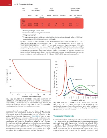
1782   Part XI  Transfusion Medicine


                          Cell          Blood              Cell                  Apparent volume
                          type       concentration       content                  of distribution

                                 Initial  End   %      Blood b  Product c  Fraction d  Liters  No. of blood
                                              change a                                 volumes
                          CD34
                          Cells
                          25 L   0.077  0.045   –45     3.8     7.9     2.3     25.9     5.3
                         15 L ×2  0.078  0.039  –50     3.9     8.1     2.3     22.2     5.1


                          a  Percentage change, end vs. initial
                          b  Estimated initial content in peripheral blood
                          c  Total product content
                          d  Total product content divided by estimated initial content in peripheral blood.  L, liters.  CD34 cell
                                                            8
                                         9
                             concentration in 10 /L, CD34 cell content x 10  cells.
                        Fig. 118.1  TWENTY HEALTHY MOBILIZED DONORS UNDERWENT EITHER A SINGLE LARGE
                        VOLUME  LEUKAPHERESIS  PROCEDURE  OF  25 L  OR  TWO  CONSECUTIVE-DAY  SMALLER
                        VOLUME PROCEDURES OF 15 L EACH. In both study groups, more than twice as many CD34 cells
                        were collected than the estimated total content of these cells in the peripheral blood before apheresis, providing
                        evidence for the existence of a large extravascular pool of peripheral blood stem cells, which becomes accessible
                        to collection by leukapheresis. (Modified from Bolan CD, Carter CS, Wesley RA, et al: Prospective evaluation of cell
                        kinetics, yields and donor experiences during a single large-volume apheresis versus two smaller volume consecutive day
                        collections of allogeneic peripheral blood stem cells. Br J Hematol 120:801, 2003, by permission of the authors.)


           100
                                                                20
                                                                15
            80                                                                  Platelets
                                                                10
          % Original component  60                              Cell number (relative scale) 9 8 7 6  Lymphocytes


            40


            20                                                   5 4 3                     Other    Granulocytes
                                                                                           mononuculear
                                                                 2                         cells
             0                                                   1
              0      0.5    1.0     1.5    2.0    2.5    3.0
                          Number of volumes exchanged            0
                                                                          0  1  2  3  4  5  6  7  8  9  10  11  12 13 14
        Fig.  118.2  RELATIONSHIP  BETWEEN  VOLUMES  REMOVED  BY
        APHERESIS AND PERCENTAGE OF THE TARGET COMPONENT                          Hemoglobin g/100 mL
        REMAINING. The relation is valid for blood volumes during red blood cell   Fig. 118.3  SCHEMATIC DISTRIBUTION OF CELLS AT THE COL-
        exchange or for plasma volumes during plasmapheresis if the target solute   LECTION  PORT  OF  A  CENTRIFUGAL  CELL  SEPARATOR.  The
        remains primarily within the intravascular compartment.   number and percentage of each cell type collected can be varied by adjusting
                                                              the  site  of  collection  along  the  interface  or  by  changing  centrifugal  force,
                                                              blood flow rate, or rate of cell removal.
        columns selectively remove LDL, very-low-density lipoprotein, and
        lipoprotein,  and  have  proved  effective  in  managing  patients  with
        homozygous hypercholesterolemia who have not responded to diet
        and cholesterol-lowering drug therapy (Fig. 118.4). A similar tech-  Therapeutic Cytapheresis
        nique, heparin-induced extracorporeal lipoprotein precipitation, uses
        low pH and negatively charged heparin to precipitate lipoproteins   Common indications for therapeutic cell removal are listed in Table
        and  remove  the  precipitate  by  filtration  online  (H.E.L.P.  Plasmat   118.1. Most of these procedures entail simple apheresis of the patient’s
        Futura, Braun Medical). Apheresis technology has also been adapted   peripheral  blood  with  diversion  for  discarding  the  affected  RBC,
        for extracorporeal phototherapy of patient leukocytes (photopheresis)   white blood cell (WBC), or platelet fraction. Special apheresis tech-
        to treat cutaneous T-cell lymphoma and to modulate the pathologic   nologies  have  been  approved,  such  as  photopheresis  devices,  that
        immune response in graft-versus-host disease (GVHD), solid organ   divert  a  cell  fraction  that  is  photochemically  treated  in  the  extra-
        transplant, and autoimmune diseases (see later discussion).  corporeal  circuit  and  returned  to  the  patient’s  circulation,  and