
Page 2066 - Hematology_ Basic Principles and Practice ( PDFDrive )
P. 2066
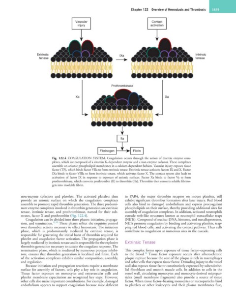
Chapter 122  Overview of Hemostasis and Thrombosis  1835


                                        Vascular                                Contact
                                         injury                                activation


                                       TF    VIIa                                   VIIIa H
                    Extrinsic           IX   X                 IXa             VIIIa L  IXa  X        Intrinsic
                     tenase                                                                           tenase


                                                           Prothrombinase


                                        Xa                      Va H                   Xa
                                                         Va L         II
                                                               Xa


                                                                IIa


                                                     Fibrinogen       Fibrin

                            Fig. 122.4  COAGULATION SYSTEM. Coagulation occurs through the action of discrete enzyme com-
                            plexes, which are composed of a vitamin K–dependent enzyme and a non-enzyme cofactor. These complexes
                            assemble on anionic phospholipid membranes in a calcium-dependent fashion. Vascular injury exposes tissue
                            factor (TF), which binds factor VIIa to form extrinsic tenase. Extrinsic tenase activates factors IX and X. Factor
                            IXa binds to factor VIIIa to form intrinsic tenase, which activates factor X. The contact system also leads to
                            activation  of  factor  IX  in  response  to  exposure  of  anionic  surfaces.  Factor  Xa  binds  to  factor Va  to  form
                            prothrombinase, which converts prothrombin (II) to thrombin (IIa). Thrombin then converts soluble fibrino-
                            gen into insoluble fibrin.


            non-enzyme  cofactors  and  platelets.  The  activated  platelets  then   in  PAR4,  the  major  thrombin  receptor  on  mouse  platelets,  still
            provide  an  anionic  surface  on  which  the  coagulation  complexes   exhibit significant thrombus formation after laser injury. Red blood
            assemble to promote rapid thrombin generation. The three predomi-  cells  also  bind  to  damaged  endothelium  and  express  procoagulant
            nant enzyme complexes involved in thrombin generation are extrinsic   phospholipids on their surface, thereby providing additional sites for
            tenase, intrinsic tenase, and prothrombinase, named for their sub-  assembly of coagulation complexes. In addition, activated neutrophils
            strates, factor X and prothrombin (Fig. 122.4).       extrude web-like structures known as neutrophil extracellular traps
              Coagulation can be divided into three phases: initiation, propaga-  (NETs). Composed of nuclear DNA, histones, and metalloproteases,
            tion, and termination. 12,13  These phases reflect the exquisite control   NETs promote coagulation by binding and activating platelets, trap-
            over thrombin activity necessary to effect hemostasis. The initiation   ping red blood cells, and activating the contact pathway. Thus cells
            phase,  which  is  predominantly  mediated  by  extrinsic  tenase,  is   contribute to coagulation at numerous sites in the cascade.
            responsible for generating the initial burst of thrombin required for
            platelet and coagulation factor activation. The propagation phase is
            largely mediated by intrinsic tenase and is responsible for the explosive   Extrinsic Tenase
            thrombin generation necessary to sustain the coagulant response. The
            termination phase, which is mediated by numerous protease inhibi-  This complex forms upon exposure of tissue factor–expressing cells
                                                                            14
            tors, ensures that thrombin generation is localized and finite. Each   to  the  blood.   Tissue  factor  exposure  occurs  after  atherosclerotic
            of the activation complexes exhibits similar composition, assembly,   plaque rupture because the core of the plaque is rich in macrophages
            and regulation.                                       and other cells that express tissue factor. Denuding injury to the vessel
              Because initiation and propagation reactions require a membrane   wall also exposes tissue factor constitutively expressed by subendothe-
            surface for assembly of factors, cells play a key role in coagulation.   lial fibroblasts and smooth muscle cells. In addition to cells in the
            Tissue  factor  exposure  on  monocytes  and  extravascular  cells  and   vessel wall, circulating monocytes and monocyte-derived micropar-
            platelet membrane capacitation are recognized key steps. However,   ticles  (small  membrane  fragments)  also  provide  a  source  of  tissue
            other cells also make important contributions. For example, damaged   factor. When tissue factor–bearing monocytes or microparticles bind
            endothelium appears to support coagulation because mice deficient   to  platelets  or  other  leukocytes  and  their  plasma  membranes  fuse,