
Page 2095 - Hematology_ Basic Principles and Practice ( PDFDrive )
P. 2095
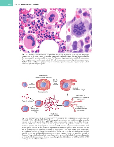
1858   Part XII  Hemostasis and Thrombosis


                       A                                                 B

                        Fig. 124.1  POLYPLOID MEGAKARYOCYTES IN THE BONE MARROW. Large polyploid megakaryo-
                        cytes are seen in the bone marrow on a typical hematoxylin- and eosin-stained slide and are recognized by
                        their abundant pink cytoplasm and large nuclei (A). The degree of polyploidization is difficult to determine.
                        Rarely, megakaryocytes can be seen in mitosis (B), and when chromosomes are aligned in metaphase plates,
                        the high ploidy level become quite apparent. In the mitotic figure illustrated, the megakaryocyte is a 16N
                        form with eight 2N metaphase plates.


                                           Production of
                                       platelet-specific granules
                                                          NF-E2
                                                          GATA1
                                  Tpo                      FOG

                               α-granules
                                                                               Cortical
                                                                               microtubule arrays
                                         Dense granules
                                                                                   Production of
                                                                               proplatelets—microtubule
                                                                                    dependent
                        Platelets release

                                                                                   Branching of
                                   Apoptosis of                                  proplatelets—actin
                                   residual cell                                    dependent
                                     body
                                                       Production
                                                      site of platelets
                        Fig. 124.2  SUMMARY OF THE MAJOR EVENTS THAT LEAD TO PLATELET FORMATION AND
                        RELEASE FROM MEGAKARYOCYTES. Hematopoietic stem cells are converted into megakaryocytes by
                        exposure to the specific growth factor TPO. TPO initiates a maturation program that amplifies the mega-
                        karyocyte  DNA  and  leads  to  synthesis  of  platelet-specific  proteins.  In  particular,  cytoskeletal  elements,
                        membrane  systems,  and  receptor  proteins  are  made  in  bulk,  and  the  megakaryocyte  becomes  filled  with
                        platelet-specific granules. Platelet production begins when microtubules aggregate in the cell cortex, and one
                        pole of the megakaryocyte spontaneously elaborates pseudopodia. These begin as large blunt pseudopodia,
                        which subsequently thin and branch into proplatelets. The branching reaction is dependent on a localized
                        assembly of actin and is inhibited by drugs that disrupt actin filaments. Platelets are assembled primarily at
                        the ends of the proplatelets. Intracellular organelles are delivered to the platelet buds along microtubule tracks
                        in the shafts. Platelets are released from the ends of proplatelets. FOG, Friend of GATA1; GATA1, GATA
                        binding protein 1; TPO, thrombopoietin.