
Page 2098 - Hematology_ Basic Principles and Practice ( PDFDrive )
P. 2098
Chapter 124 Megakaryocyte and Platelet Structure 1861
the maturation of hematopoietic stem cells into proplatelet-producing tubulin as a downstream effector of the megakaryocyte transcription
megakaryocytes in the second half. Human embryonic stem cells can factor NF-E2 that is absent in NF-E2–deficient megakaryocytes.
3
be coaxed to differentiate into mature megakaryocytes, although the Second, genetic elimination of the β 1-tubulin gene in mice results in
6
process takes several more days in culture. Recently, platelets have thrombocytopenia. Third, megakaryocytes isolated from β 1 -tubulin
been generated from induced human pluripotent stem cells in culture knockout mice fail to form proplatelets in vitro and instead extend
using a doxycycline-controlled c-MYC expression vector. 4 only a small number of blunt protrusions.
The first event that signals proplatelet production is the consolida-
tion of microtubules into large bundles at the megakaryocyte cortex
PLATELET FORMATION that subsequently are reorganized into parallel bundles in the shafts
8
of the proplatelets (Fig. 124.6). Microtubule bundles are thick near
Proplatelets and the Cytoskeletal the body of the megakaryocyte as they enter the proplatelet shaft but
become progressively thinner along the shaft, such that only 5–10
Mechanics of Platelet Formation microtubules remain at the end of the proplatelet. Of note, the
microtubule bundles that run down the proplatelet shaft make
The discovery of TPO and the development of megakaryocyte cul- characteristic U turns in the tips and reenter the shaft, forming
tures that reconstitute platelet formation in vitro have allowed visu- teardrop-shaped structures (Fig. 124.7). This creates a bipolar orien-
5
alization of megakaryocytes in the act of forming platelets. The tation of bundles near the proplatelet tip, a geometry required to
actual mechanical process of platelet production begins when mature explain the bidirectional granule and organelle traffic observed in
megakaryocytes start to elaborate proplatelets (see Fig. 124.2 and Fig. proplatelets. The looped arrangement of microtubules in proplatelet
124.5). This process is distinguished by the erosion of one pole of tips also constrains the elongation mechanism used to grow proplate-
the megakaryocyte cytoplasm (see Fig. 124.5). Multiple thick pseu- lets because of an insufficient number of free microtubule ends to
dopodia are extended and subsequently elongate to yield thin tubules. nucleate this reaction.
As these slender tubules grow, they branch repeatedly and develop Direct visualization of microtubule dynamics in living megakaryo-
periodic densities along their length that impart a beaded appear- cytes using green fluorescent protein (GFP) technology has provided
5,6
ance. The first insight into the cytoskeletal mechanics of platelet insights into how microtubules orient to power proplatelet elongation
formation dates from the work of Tablin and colleagues, who showed (see Fig. 124.4A,B). End-binding protein 3 (EB3), a microtubule
that proplatelet formation is dependent on microtubules; that is, plus end-binding protein associated only with growing microtubules,
proplatelet elaboration is inhibited by microtubule poisons. Micro- fused to GFP was retrovirally expressed in murine megakaryocytes
tubule poisons are effective because the extension of proplatelets from and used as a marker to localize microtubule plus ends and to follow
7
the megakaryocyte is mediated by the assembly of microtubules and plus end dynamics. Immature megakaryocytes without proplatelets
their reorganization into cortical bundles. Cortical bundles align in use a centrosomal-coupled microtubule nucleation/assembly reac-
the shafts of proplatelets, and proplatelet elongation is driven by tion, which appears as a prominent starburst pattern when visualized
sliding movements between overlapping microtubules composed of with EB3-GFP. Microtubules assemble only from the centrosome and
7
these bundles. The microtubule bundles form loops at the end of grow outward into the cell cortex, where they turn and run parallel
each proplatelet, and ultimately a single microtubule is rolled into a to the cell edges. Just before proplatelet production begins, however,
coil at the proplatelet end to define the platelet territory. Cytoplasmic centrosomal assembly ends and microtubules release and consolidate
tubulin in solution is an αβ dimer that reversibly polymerizes into into the cortex as bundles. Fluorescence time-lapse microscopy of
microtubules, which are long, hollow cylinders with an outer diam- living, proplatelet-producing megakaryocytes expressing EB3-GFP
eter of 25 nm. Several studies reveal an essential role in platelet reveals that as proplatelets elongate, microtubules assemble continu-
biogenesis for β 1 tubulin, a divergent and lineage-specific β tubulin ously throughout the entire proplatelet. EB3-GFP studies also reveal
that is a major component of the megakaryocyte proplatelet cytoskel- that microtubules polymerize in both directions in proplatelets—that
eton and marginal microtubule coil of the platelet. β 1 Tubulin, which is, toward both the tips and cell body—demonstrating that microtu-
is expressed exclusively in platelets and megakaryocytes during the bules composing the bundles have a mixed polarity. The cytoplasmic
late stages of megakaryocyte development, is essential for the produc- Ran-binding protein, RanBP10, is a β 1 -tubulin–binding protein that
9
tion of normal numbers of platelets, as well as for the discoid shape appears to regulate the assembly of proplatelet microtubules. Even
of platelets. The evidence supporting the role of β 1 tubulin in these though microtubules are continuously assembling at their plus ends
processes comes from several sources. First, mRNA subtraction in proplatelets, polymerization per se does not provide the force for
between wild-type and NF-E2–deficient megakaryocytes identifies β 1 proplatelet elongation. First, the rates of microtubule polymerization
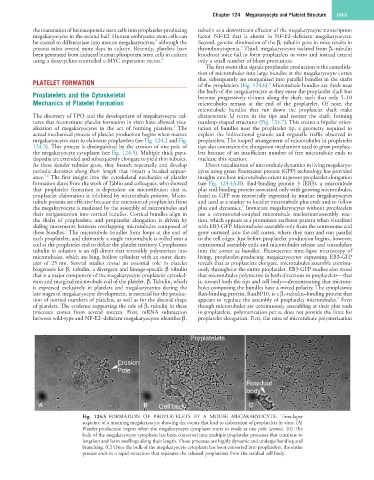
Fig. 124.5 FORMATION OF PROPLATELETS BY A MOUSE MEGAKARYOCYTE. Time-lapse
sequence of a maturing megakaryocyte showing the events that lead to elaboration of proplatelets in vitro. (A)
Platelet production begins when the megakaryocyte cytoplasm starts to erode at one pole (arrow). (B) The
bulk of the megakaryocyte cytoplasm has been converted into multiple proplatelet processes that continue to
lengthen and form swellings along their length. These processes are highly dynamic and undergo bending and
branching. (C) Once the bulk of the megakaryocyte cytoplasm has been converted into proplatelets, the entire
process ends in a rapid retraction that separates the released proplatelets from the residual cell body.