
Page 2097 - Hematology_ Basic Principles and Practice ( PDFDrive )
P. 2097
1860 Part XII Hemostasis and Thrombosis
PP
CB
A
Distance
0 (µm) 10
0
Time
(sec)
C
B 60
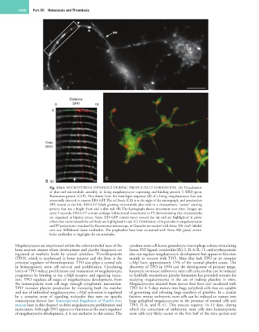
Fig. 124.4 MICROTUBULE DYNAMICS DURING PROPLATELET FORMATION. (A) Visualization
of plus-end microtubule assembly in living megakaryocytes expressing end-binding protein 3 (EB3)-green
fluorescent protein (GFP). First frame from the time-lapse sequence (B) of a living megakaryocyte that was
retrovirally directed to express EB3-GFP. The cell body (CB) is at the right of the micrograph, and proplatelets
(PP) extend to the left. EB3-GFP labels growing microtubule plus ends in a characteristic “comet” staining
pattern that has a bright front and a dim tail. (B) The kymograph shows movement over time. Images are
every 5 seconds. EB3-GFP comets undergo bidirectional movements in PP, demonstrating that microtubules
are organized as bipolar arrays. Some EB3-GFP comets move toward the tip and are highlighted in green;
others that move toward the cell body are highlighted in red. (C) Distribution of α-granules in megakaryocytes
and PP projections visualized by fluorescence microscopy. α-Granules are stained with Alexa 568 (red)–labeled
anti–von Willebrand factor antibodies. The proplatelets have been co-stained with Alexa 488 (green) antitu-
bulin antibodies to highlight the microtubules.
Megakaryocytes are imprisoned within the subendothelial layer of the cytokine stem cell factor, granulocyte-macrophage colony-stimulating
bone marrow sinuses where development and platelet biogenesis are factor, FLT ligand, interleukin (IL)-3, IL-6, IL-11, and erythropoietin
regulated at multiple levels by several cytokines. Thrombopoietin also can regulate megakaryocyte development but appear to function
(TPO), which is synthesized in bone marrow and the liver, is the mainly in concert with TPO. Mice that lack TPO or its receptor
principal regulator of thrombopoiesis. TPO also plays a central role c-Mpl have approximately 15% of the normal platelet count. The
in hematopoietic stem cell survival and proliferation. Circulating discovery of TPO in 1994 and the development of primary mega-
levels of TPO induce proliferation and maturation of megakaryocyte karyocyte or mouse embryonic stem cell cultures that can be induced
progenitors by binding to the c-Mpl receptor and signaling induc- to faithfully reconstitute platelet formation has provided systems for
tion. TPO regulates all stages of megakaryocyte development, from studying megakaryocytes in the act of making platelets in vitro.
the hematopoietic stem cell stage through cytoplasmic maturation. Megakaryocytes isolated from mouse fetal liver and incubated with
TPO increases platelet production by increasing both the number TPO for 4–5 days mature into huge polyploid cells that are capable
and size of individual megakaryocytes. c-Mpl activation is regulated of generating and releasing large numbers of platelets. In a similar
by a complex array of signaling molecules that turn on specific fashion, mouse embryonic stem cells can be induced to mature into
transcription factors (see Transcriptional Regulation of Platelet For- large polyploid megakaryocytes in the presence of stromal cells and
mation later in this chapter) to drive megakaryocyte proliferation and TPO, IL-6, and IL-11. This process requires 10–12 days, during
maturation. Although TPO appears to function as the main regulator which the conversion of embryonic stem cells into hematopoietic
of megakaryocyte development, it is not exclusive in this action. The stem cells very likely occurs in the first half of the time period and