
Page 2100 - Hematology_ Basic Principles and Practice ( PDFDrive )
P. 2100
Chapter 124 Megakaryocyte and Platelet Structure 1863
contrast, mice lacking the actin turnover protein ADF have normal
−/−
34
platelet counts and morphology. On the other hand, when ADF
mice are crossed with cofilin-1 knockout mice, platelet production is
severely reduced and morphologies of platelets are highly variable.
Mice that contain megakaryocytes that specifically lack profilin 1, a
small protein that promotes actin filament assembly, have macro-
35
thrombocytopenia with reduced platelet counts. Profilin-null
platelets have a thickened microtubule coil with hyperacetylated
microtubules, and in some ways, the profilin 1 knockout phenotype
is similar to the behavior of platelets in Wiskott-Aldrich syndrome,
or in WASp knockout mice. Defective proplatelet production has also
been observed in mice in which the small regulatory GTPases Rho,
Cdc42, and Rac have been deleted in the megakaryocyte lineage. 36,37
In addition to playing an essential role in proplatelet elongation,
the microtubules lining the shafts of proplatelets serve a secondary
function: transport of membrane, organelles, and granules into pro-
A platelets and assembling platelets at proplatelet ends (see Fig. 124.4C).
Organelles are sent individually from the cell body into the proplate-
lets, where they move bidirectionally until they are captured at pro-
14
platelet tips. Immunofluorescence and electron microscopic studies
indicate that organelles are intimately associated with microtubules,
and actin poisons do not diminish organelle motion. Thus movement
appears to involve microtubule-based forces. Bidirectional organelle
movement is conveyed in part by the bipolar arrangement of micro-
tubules within the proplatelet because kinesin-coated latex beads
move in both directions over the microtubule arrays of permeabilized
proplatelets. Of the two major microtubule motors, kinesin and
cytoplasmic dynein, only the plus end-directed kinesin is localized in
a pattern similar to organelles and granules, and is likely responsible
for transporting these elements along microtubules. It appears that
two mechanisms of organelle and granule movement are involved in
platelet assembly: first, organelles and granules travel along microtu-
bules, and second, the microtubules themselves slide bidirectionally
in relation to other motile filaments, indirectly moving organelles
along proplatelets in a piggyback manner.
B Although the roles of microtubules and actin filaments in pro-
platelet development have been extensively studied, our understand-
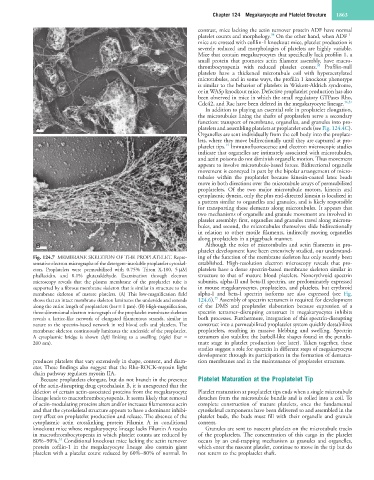
Fig. 124.7 MEMBRANE SKELETON OF THE PROPLATELET. Repre- ing of the function of the membrane skeleton has only recently been
sentative electron micrographs of the detergent-insoluble proplatelet cytoskel- established. High-resolution electron microscopy reveals that pro-
eton. Proplatelets were permeabilized with 0.75% Triton X-100, 5 µM platelets have a dense spectrin-based membrane skeleton similar in
phallacidin, and 0.1% glutaraldehyde. Examination through electron structure to that of mature blood platelets. Nonerythroid spectrin
microscopy reveals that the plasma membrane of the proplatelet tube is subunits, alpha-II and beta-II spectrin, are predominately expressed
supported by a fibrous membrane skeleton that is similar in structure to the in mouse megakaryocytes, proplatelets, and platelets, but erythroid
membrane skeleton of mature platelets. (A) This low-magnification field alpha-I and beta-I spectrin isoforms are also expressed (see Fig.
15
shows that an intact membrane skeleton laminates the underside and extends 124.6). Assembly of spectrin tetramers is required for development
along the entire length of proplatelets (bar = 1 µm). (B) High-magnification, of the DMS and proplatelet elaboration because expression of a
three-dimensional electron micrograph of the proplatelet membrane skeleton spectrin tetramer–disrupting construct in megakaryocytes inhibits
reveals a lattice-like network of elongated filamentous strands, similar in both processes. Furthermore, integration of this spectrin-disrupting
nature to the spectrin-based network in red blood cells and platelets. The construct into a permeabilized proplatelet system quickly destabilizes
membrane skeleton continuously laminates the underside of the proplatelet. proplatelets, resulting in massive blebbing and swelling. Spectrin
A cytoplasmic bridge is shown (left) linking to a swelling (right) (bar = tetramers also stabilize the barbell-like shapes found in the penulti-
200 nm). mate stage in platelet production (see later). Taken together, these
studies suggest a role for spectrin in different steps of megakaryocyte
development through its participation in the formation of demarca-
produces platelets that vary extensively in shape, content, and diam- tion membranes and in the maintenance of proplatelet structure.
eter. These findings also suggest that the Rho-ROCK-myosin light
chain pathway regulates myosin IIA.
Because proplatelets elongate, but do not branch in the presence Platelet Maturation at the Proplatelet Tip
of the actin-disrupting drug cytochalasin B, it is unexpected that the
deletion of certain actin-associated proteins from the megakaryocyte Platelet maturation at proplatelet tips ends when a single microtubule
lineage leads to macrothrombocytopenia. It seems likely that removal detaches from the microtubule bundle and is rolled into a coil. To
of actin-modulating proteins alters and/or increases filamentous actin complete construction of mature platelets, once the fundamental
and that the cytoskeletal structure appears to have a dominant inhibi- cytoskeletal components have been delivered to and assembled in the
tory effect on proplatelet production and release. The absence of the platelet buds, the buds must fill with their organelle and granule
cytoplasmic actin crosslinking protein Filamin A in conditional content.
knockout mice whose megakaryocyte lineage lacks Filamin A results Granules are sent to nascent platelets on the microtubule tracks
in macrothrombocytopenia in which platelet counts are reduced by of the proplatelets. The concentration of this cargo in the platelet
33
80%–90%. Conditional knockout mice lacking the actin turnover occurs by an end-trapping mechanism as granules and organelles,
protein cofilin-1 in the megakaryocyte lineage also contain giant which enter the nascent platelet, continue to move in the tip but do
platelets with a platelet count reduced by 60%–80% of normal. In not return to the proplatelet shaft.