
Page 2142 - Hematology_ Basic Principles and Practice ( PDFDrive )
P. 2142
1 2
X
IXα
3 4
ATIII ATIII
VIIa IIa Xa IIa IIa
Va VIIIa
Va
TF HS HS TM TM
Xa IXa PC
TF•VIIa Xa•Va VIIIa•IXa HS•ATIII•IIa HS•ATIII•Xa TM•IIa TM•IIa•PC
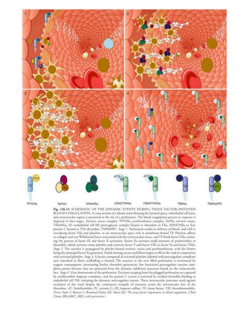
Fig. 126.15 SCHEMATIC OF THE DYNAMIC EVENTS DURING TISSUE FACTOR–INITIATED
BLOOD COAGULATION. A cross-section of a blood vessel showing the luminal space, endothelial cell layer,
and extravascular region is presented at the site of a perforation. The blood coagulation process in response is
depicted in four stages. Extrinsic tenase complex, TF•VIIa; prothrombinase complex, Xa•Va; intrinsic tenase,
VIIIa•IXa; AT–endothelial cell HS proteoglycan complex bound to thrombin or FXa, HS•AT•(IIa or Xa);
protein C bound to TM–thrombin, TM•IIa•PC. Stage 1. Perforation results in delivery of blood, and with it
circulating factor VIIa and platelets, to an extravascular space rich in membrane-bound TF. Platelets adhere
to collagen and von Willebrand factor associated with the extravascular tissue, and TF binds factor VIIa, initiat-
ing the process of factor IX and factor X activation. Factor Xa activates small amounts of prothrombin to
thrombin, which activates more platelets and converts factor V and factor VIII to factor Va and factor VIIIa.
Stage 2. The reaction is propagated by platelet-bound intrinsic tenase and prothrombinase, with the former
being the principal factor Xa generator. Initial clotting occurs and fibrin begins to fill in the void in cooperation
with activated platelets. Stage 3. A barrier composed of activated platelets ladened with procoagulant complexes
and enmeshed in fibrin scaffolding is formed. The reaction in the now filled perforation is terminated by
reagent consumption, attenuating further thrombin generation, but functional procoagulant enzyme com-
plexes persist because they are protected from the dynamic inhibitory processes found on the intravascular
face. Stage 4. View downstream of the perforation. Enzymes escaping from the plugged perforation are captured
by antithrombin–heparan complexes, and the protein C system is activated by residual thrombin binding to
endothelial cell TM, initiating the dynamic anticoagulant system. These intravascular processes work against
occlusion of the vessel despite the continuous resupply of reactants across the intravascular face of the
thrombus. AT, Antithrombin; PC, protein C; HS, heparan sulfate; TF, tissue factor; TM, thrombomodulin.
(From Orfeo T, Butenas S, Brummel-Ziedins KE, Mann KG: The tissue factor requirement in blood coagulation. J Biol
Chem 280:42887, 2005, with permission.)