
Page 213 - Hematology_ Basic Principles and Practice ( PDFDrive )
P. 213
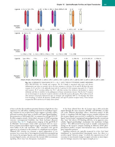
Chapter 16  Cytokine/Receptor Families and Signal Transduction  165


                 Ligands  IL-1α / IL-1β/IL-1Ra  IL-33  IL-36α / IL-36β / IL-36γ/IL-36Ra/ IL-38  IL-18  IL-37


                Receptor
                  chains
                          IL-1R1/ IL-1R3  IL-1R4/ IL-1R3  IL-36R/ IL-1R3  IL-1R5/ IL-1R7  IL-1R5

                 Ligands  IL-17A/ IL-17F  IL-17C     ?        IL-17E(IL-25)  IL-17B


                Receptor
                  chains
                       IL-17RA/ IL-17RC     IL-17A/ IL-17RE  IL-17RA/ IL-17RD       IL-17RA/ IL-17RB       ? / IL-17RB
                 Ligands  Type I IFN’ s  IFN-γ  IL-10     IL-22       IL-26   IL-28/IL-29  IL-10/ IL-20/IL-24  IL-20/IL-24


                Receptor
                  chains
                       IFNAR1/IFNAR2  IFN-γR1/IFN-γR2  IL-10R1/IL-10R2  IL-22R1/IL-10R2  IL-20R1/IL-10R2  IL-28R1/IL-10R2  IL-20R1/IL-20R2  IL-22R1-IL-20R2
                            Fig. 16.2  SCHEMATIC DIAGRAMS OF IL-1, IL-17, AND TYPE II CYTOKINE FAMILY MEMBERS
                            AND  RECEPTORS.  IL-1  family  and  receptors.  The  IL-1  family  is  associated  with  acute  and  chronic
                            inflammation and plays an essential role in the host response to infection. IL-1α and IL-1β share the same
                            receptor. IL-33 and the IL-36 subfamily share with IL-1 and the IL-1R3 receptors (top panel); IL-17 family
                            and  receptors.  IL-17  secretion  defines  the Th17  cells  that  mediate  host  defensive  mechanisms  to  various
                            infections and that are involved in the pathogenesis of many autoimmune diseases. The five IL-17 receptors
                            are not homologous to any known receptors and show considerable sequence divergence. They harbor extracel-
                            lular domains composed of fibronectin type III domains and cytoplasmic SEF–IL-17R domains that show
                            loose homology to toll–IL-1R domains (middle panel); and type II cytokine receptor family. These include
                            receptors for IFNs and the IL-10 family (lower panel).


            is bent such that the membrane-proximal domains of gp130 are close   It  has  been  debated  how  the  16  human  type  I  IFN  molecules
            together at the cell surface, enabling activation of intracellular signal-  signal through the same receptors, IFNAR1 and IFNAR2, yet they
            ing. Variation in the receptor bend angles suggests a possible confor-  can  evoke  different  physiologic  effects.  Structural  analysis  of  this
                                                              8
            mational transition from open to closed states upon ligand binding.    family in complexes with this single receptor complex indicates that
            Reconstruction of full-length JAK1 in conjunction with gp130/IL-6/  the receptor-ligand cross-reactivity is enabled by conserved receptor-
            IL-6Rα complex reveals a three-lobed structure of JAK1 possessing   ligand “anchor points” interspersed among ligand-specific interactions
            extensive  intersegmental  flexibility  that  likely  facilitates  allosteric   that “tune” the relative IFN-binding affinities, in an apparent extra-
                            9
            activation  (Fig.  16.7).   Single-particle  imaging  of  the  gp130/IL-6/  cellular  “ligand  proofreading”  mechanism  that  modulates  biologic
                                                                       10
            IL-6Rα/Jak1  holocomplex  shows  JAK1  associated  with  the  mem-  activity.  This differential binding leads to variable conformational
            brane proximal intracellular regions of gp130, abutting the would-be   change in the receptor complex resulting in different STAT phos-
            inner  leaflet  of  the  cell  membrane.  JAK1  association  with  gp130   phorylation profiles, receptor internalization rates, and downstream
            appears to be enhanced by the presence of a membrane environment.   gene expression patterns.
            Mutated  JAK  proteins  can  transmit  a  signal  independent  of  the   Signaling  networks  are  typically  measured  in  either  their  basal
            receptor, but a recent line of investigation revealed that manipulating   (minimum)  or  hyperstimulated  (maximum)  states,  but  there  is  a
            the receptor by surrogate ligands, such as diabodies to mimic dimeric   cytokine signal “dynamic range,” in which the responsiveness of cell
            receptor-ligand system, can attenuate the mutant JAK2 downstream   outcomes to incremental changes in signal activation is more impor-
            signaling, which may have therapeutic implications.   tant for biologic outcome than signal strength per se. 11