
Page 2261 - Hematology_ Basic Principles and Practice ( PDFDrive )
P. 2261
2008 Part XII Hemostasis and Thrombosis
mild deficiency state of all of the vitamin K–dependent proteins is
an inherited defect in the γ-carboxylase enzyme required for the
posttranslational modification of these proteins.
CLINICAL FEATURES OF HEMOPHILIA
The clinical symptoms and signs of hemophilia A and B are essentially
identical and relate to the propensity for prolonged and excessive
bleeding. The bleeding tendency in hemophilia is determined in large
part by the baseline activity level of the deficient or defective clotting
factor. Thus in severe hemophilia (A and B), in which the baseline
activity level of clotting factor is below 1% (0.01 IU/mL), spontane-
ous bleeding usually occurs multiple times each year. With a moderate
factor deficiency state of 1% to 5% (0.01–0.05 IU/mL), spontaneous
bleeding is typically infrequent, but excessive and prolonged bleeding
can occur with trauma and invasive surgical or dental procedures.
Finally, in mild disease, with factor levels of 5% to 40% (0.05–
0.40 IU/ml), excessive bleeding is usually only documented with
trauma or invasive procedures.
Pathologic bleeding can occur in the neonatal period, when intra-
cranial bleeding can develop after traumatic delivery, especially in
infants with severe hemophilia. However, most frequently, severe
disease manifests with easy bruising or soft tissue or joint bleeding
between 6 and 18 months of age when the young child becomes more
mobile. In mild hemophilia, the disease may remain silent for many
years, and occasionally a new diagnosis of hemophilia may be made
in those older than 60 years of age when challenged with a surgical
procedure.
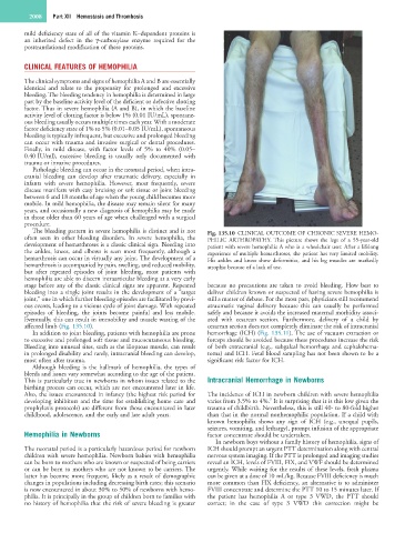
The bleeding pattern in severe hemophilia is distinct and is not Fig. 135.10 CLINICAL OUTCOME OF CHRONIC SEVERE HEMO-
often seen in other bleeding disorders. In severe hemophilia, the PHILIC ARTHROPATHY. This picture shows the legs of a 55-year-old
development of hemarthroses is a classic clinical sign. Bleeding into patient with severe hemophilia A who is a wheelchair user. After a lifelong
the ankles, knees, and elbows is seen most frequently, although a experience of multiple hemarthroses, the patient has very limited mobility.
hemarthrosis can occur in virtually any joint. The development of a His ankles and knees show deformities, and his leg muscles are markedly
hemarthrosis is accompanied by pain, swelling, and reduced mobility, atrophic because of a lack of use.
but after repeated episodes of joint bleeding, most patients with
hemophilia are able to discern intraarticular bleeding at a very early
stage before any of the classic clinical signs are apparent. Repeated because no precautions are taken to avoid bleeding. How best to
bleeding into a single joint results in the development of a “target deliver children known or suspected of having severe hemophilia is
joint,” one in which further bleeding episodes are facilitated by previ- still a matter of debate. For the most part, physicians still recommend
ous events, leading to a vicious cycle of joint damage. With repeated atraumatic vaginal delivery because this can usually be performed
episodes of bleeding, the joints become painful and less mobile. safely and because it avoids the increased maternal morbidity associ-
Eventually, this can result in immobility and muscle wasting of the ated with cesarean section. Furthermore, delivery of a child by
affected limb (Fig. 135.10). cesarean section does not completely eliminate the risk of intracranial
In addition to joint bleeding, patients with hemophilia are prone hemorrhage (ICH) (Fig. 135.11). The use of vacuum extraction or
to excessive and prolonged soft tissue and mucocutaneous bleeding. forceps should be avoided because these procedures increase the risk
Bleeding into unusual sites, such as the iliopsoas muscle, can result of both extracranial (e.g., subgaleal hemorrhage and cephalohema-
in prolonged disability and rarely, intracranial bleeding can develop, toma) and ICH. Fetal blood sampling has not been shown to be a
most often after trauma. significant risk factor for ICH.
Although bleeding is the hallmark of hemophilia, the types of
bleeds and issues vary somewhat according to the age of the patient.
This is particularly true in newborns in whom issues related to the Intracranial Hemorrhage in Newborns
birthing process can occur, which are not encountered later in life.
Also, the issues encountered in infancy (the highest risk period for The incidence of ICH in newborn children with severe hemophilia
9
developing inhibitors and the time for establishing home care and varies from 3.5% to 4%. It is surprising that it is this low given the
prophylaxis protocols) are different from those encountered in later trauma of childbirth. Nevertheless, this is still 40- to 80-fold higher
childhood, adolescence, and the early and late adult years. than that in the normal nonhemophilic population. If a child with
known hemophilia shows any sign of ICH (e.g., unequal pupils,
seizures, vomiting, and lethargy), prompt infusion of the appropriate
Hemophilia in Newborns factor concentrate should be undertaken.
In newborn boys without a family history of hemophilia, signs of
The neonatal period is a particularly hazardous period for newborn ICH should prompt an urgent PTT determination along with central
children with severe hemophilia. Newborn babies with hemophilia nervous system imaging. If the PTT is prolonged and imaging studies
can be born to mothers who are known or suspected of being carriers reveal an ICH, levels of FVIII, FIX, and VWF should be determined
or can be born to mothers who are not known to be carriers. The urgently. While waiting for the results of these levels, fresh plasma
latter has become more frequent, likely as a result of demographic can be given at a dose of 10 mL/kg. Because FVIII deficiency is much
changes in populations including decreasing birth rates; this scenario more common than FIX deficiency, an alternative is to administer
is now encountered in about 30% to 50% of newborns with hemo- FVIII concentrate and determine the PTT 10 to 15 minutes later. If
philia. It is principally in the group of children born to families with the patient has hemophilia A or type 3 VWD, the PTT should
no history of hemophilia that the risk of severe bleeding is greater correct; in the case of type 3 VWD this correction might be