
Page 2263 - Hematology_ Basic Principles and Practice ( PDFDrive )
P. 2263
2010 Part XII Hemostasis and Thrombosis
and symptoms of chronic hemophilic arthropathy while other imperceptible, particularly in very young children who may not
patients may not develop joint damage despite repeated joint bleeds. vocalize the occurrence of trauma. Mild trauma may even occur while
the child is asleep, causing the child to wake up with joint pain and
swelling. The onset of a hemarthrosis is often signaled by a feeling of
Soft Tissue Hemorrhages and Muscle Bleeds warmth and tingling in the joint. This may last for several hours
before increasing pain and limitation of joint movement set in. In
Bleeding into soft tissues includes spontaneous and trauma-related patients with severe hemophilia, bleeding into the joint will continue
bleeding into subcutaneous tissues and muscles. Superficial hemato- until the patient receives hemostatic therapy or the pressure within
mas (bruises) may resolve spontaneously without the need for treat- the joint increases to the point of causing occlusion of bleeding vessels
ment, and as such, bruising is not an indication for clotting factor and bleeding cessation. In the later scenario, the pain in the joint may
replacement. However, in moderate and severe hemophilia, soft tissue be excruciating. In patients with moderate or mild hemophilia, the
hematomas often undergo progressive enlargement and may need to bleeding may stop without the administration of hemostatic replace-
be treated. Furthermore, some soft tissue bleeds (e.g., retroperitoneal ment but only after substantial bleeding has occurred. Consequently,
bleeds or hematomas of the neck) can cause extensive blood loss and joint bleeds should be treated as soon as possible to minimize the
be life or organ threatening because of their propensity to expand, extent of bleeding and reduce the risk of long-term disabling sequelae.
thus causing compression of adjacent organs, blood vessels, and Blood in a joint leads to joint damage through at least three
nerves and the airway in the case of a neck hematoma. mechanisms: iron toxicity, inflammation, and mechanical distension
Muscle bleeds are quite common in hemophilia. The muscles of the joint. Repeated joint bleeds may result in inflammation and
most often involved are, in descending order of frequency, the calf, hyperplasia of the synovial tissue within the joint (a situation referred
12
thigh, buttocks, and forearm. Bleeds into these locations can lead to to as synovitis). Synovitis is the first step toward the development of
compartment syndrome, which is an emergency situation (see box hemophilic arthropathy. Any joint that undergoes repeated bleeds is
on Compartment Syndrome). A particularly problematic muscle referred to as a target joint. The strict definition of a target joint is
bleed is a bleed into the iliopsoas muscle, a large muscle in the hip still under discussion, but in general, most clinicians tend to accept
region. Such bleeds can rapidly expand because there is no surround- that a joint that bleeds three or four times in a 6-month period
ing connective tissue to restrict their growth. Consequently, significant qualifies as a target joint. 13
bleeding can occur into this muscle, potentially leading to the need A target joint is a manifestation of synovitis, and if not managed
for blood transfusion. Patients with iliopsoas muscle bleeds have pain with long-term prophylaxis, such a joint will continue to bleed and
and restriction of movement around the hip joint; they tend to will ultimately become a chronically damaged, arthropathic joint.
maintain the leg in a flexed position. Because of increased pressure Early stage hemophilic arthropathy is characterized by synovial
on the femoral nerve, they may complain of paresthesia, hyperesthe- hyperplasia, extensive destruction of articular cartilage, progressive
sia, or weakness of the quadriceps muscle. An iliopsoas bleed can be loss of joint space, cystic changes within the subchondral bone,
confused with a hemarthrosis of the hip (see box on Hip Joint osteoporosis, and atrophy of surrounding muscles. The final stage of
Bleeds). Urgent ultrasonography or magnetic reso nance imaging hemophilic arthropathy is a deformed and dysfunctional joint. At this
(MRI) examination is required to differentiate between the two stage, joint bleeds become less frequent as synovial hypertrophy
conditions. Iliopsoas muscle bleeds need prolonged treatment with becomes less prominent.
factor concentrates, as well as immobilization and physical therapy, Determining the status of joints of patients with hemophilia at a
and consequently they usually necessitate hospitalization (see box on given time or longitudinally over time necessitates both clinical and
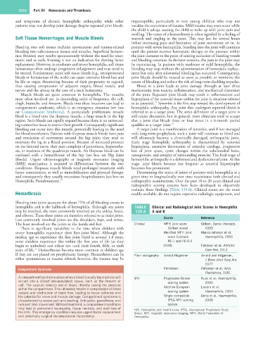
Hemophilic Pseudotumors). 10 radiographic examinations. Over the past 10 to 20 years clinical and
radiographic scoring systems have been developed to objectively
evaluate these findings (Table 135.6). Clinical scores are the most
Hemarthrosis readily available, do not require expensive radiologic equipment, and
Bleeding into joints accounts for about 75% of all bleeding events in
hemophilia and is the hallmark of hemophilia. Although any joints TABLE Clinical and Radiological Joint Scores in Hemophilia
may be involved, the most commonly involved are the ankles, knees, 135.6 A and B
and elbows. These three joints are therefore referred to as index joints.
Less commonly involved joints are the shoulders, hips, and wrists. Scores Reference
The least involved are the joints in the hands and feet. Clinical WFH joint score Gilbert, Semin Hematol,
There is significant variability in the time when children with (Gilbert score) 1993
severe hemophilia experience their first joint bleed. Although the Modified WFH joint Manco-Johnson et al,
median age to experience the first joint bleed is around 1.8 years, score Colorado Haemophilia, 2000
some children experience this within the first year of life (as they PE-1 and PE-0.5
begin to ambulate) and others not until their fourth, fifth, or sixth HJHS Feldman et al, Arthritis
11
years of life. Hemarthroses become more common as children age Care Res, 2011
if they are not placed on prophylactic therapy. Hemarthrosis can be Plain radiography Arnold-Hilgartner Arnold and Hilgartner,
either spontaneous or trauma related; however, the trauma may be J Bone Joint Surg Am,
1977
Compartment Syndrome Pettersson Petterson et al, Acta
Paediatrica, 1981
A compartment syndrome arises when a bleed (usually trauma induced) MRI Progressive Denver Nuss et al, Haemophilia,
occurs into a closed (encapsulated) space, such as the forearm or scoring system 2000
calf. The capsule restricts exit of blood, thereby raising the pressure
within the compartment. This ultimately results in compression of blood Additive European Lundin et al,
vessels and obstruction of blood flow, leading to tissue ischemia and scoring system Haemophilia, 2004
the potential for nerve and muscle damage. Compartment syndrome is Single compatible Doria et al, Haemophilia,
characterized by severe pain and swelling, limb pallor, paresthesia, and IPSG MRI scoring 2008
reduced limb movement. Without treatment, a compartment syndrome system
may lead to permanent neuropathy, tissue necrosis, and even loss of HJS, Hemophilia joint health score; IPSG, International Prophylaxis Study
the limb. This emergency condition requires urgent factor replacement Group; MRI, magnetic resonance imaging; WFH, World Federation of
and potentially surgical decompression (fasciotomy). Hemophilia.