
Page 2262 - Hematology_ Basic Principles and Practice ( PDFDrive )
P. 2262
Chapter 135 Hemophilia A and B 2009
TABLE Reasons for Differences in Bleeding Phenotype
135.5
1. Differences in factor levels
2. Differences in mutations causing hemophilia: null versus non-null
mutations
3. Coinheritance of other bleeding disorders
• VWD
• mild functional platelet disorders
4. Coinheritance of prothrombotic disorders
• factor V Leiden
• prothrombin G20210A mutation
• low levels of protein C, protein S, or antithrombin
5. Differences in the pharmacokinetic handling of factor that might be
associated with patients’ ABO blood group and endogenous levels of
VWF in the case of hemophilia A
6. Differences in levels of physical activity
7. Differences in the structural integrity of joints, making patients
more or less susceptible to joint bleeds and joint damage.
VWD, von Willebrand disease; VWF, von Willebrand factor.
Bleeding in hemophilia is broadly correlated with the endogenous
level of clotting factor. Patients with severe forms of hemophilia
defined as having an endogenous FVIII or FIX level of less than 1%
(<0.01 IU/mL) will develop spontaneous bleeds throughout their
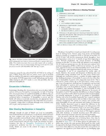
Fig. 135.11 INTRACRANIAL BLEEDING IN HEMOPHILIA. A com- lives beginning at about 6 to 12 months of age when beginning to
puted tomography scan shows an intracranial bleed in a person with hemo- crawl. Without prophylaxis, the annual incidence of bleeding
philia. This complication most often occurs after trauma with an incidence increases in the first 5–6 years of life and plateaus at an average of
of five per 1000 per year in patients younger than age 5 years and 1% to 2% 20–30 bleeds per patient per year. For the most part, patients with
per year in persons with hemophilia older than age 55 years. moderate hemophilia (FVIII or FIX level of between 1% and 5%
[0.01 and 0.05 IU/mL]) and mild hemophilia (factor levels of >5%
[>0.05 IU/mL]) only experience bleeding with trauma or surgery.
Hemophilia A and B are characterized by similar types of
temporary caused by the reduced half-life of FVIII in the setting of bleeds. However, the severity and frequency of bleeding may vary
type 3 VWD. If the PTT does not correct, then it can be assumed considerably among individuals with the same factor activity level.
that the child has FIX deficiency, and an appropriate FIX concentrate Although not conclusively proven, a number of studies suggest that
can be given. If FVIII concentrate is used, fresh plasma can be given the bleeding phenotype in hemophilia B seems to be less severe than
while waiting for the PTT results to cover the possibility that the that in hemophilia A patients with comparable factor levels. This is
child has hemophilia B. reflected in a lower median age at first joint bleed, a lower median
age at start of prophylaxis, increased use of prophylaxis, and increased
joint arthroplasty in patients with severe hemophilia A compared
Circumcision in Newborns with those with severe hemophilia B. Similarly, a higher proportion
of children with moderate hemophilia A than hemophilia B tend
Surprisingly, bleeding after circumcision only occurs in about half of to be on prophylaxis, again suggesting differences in the severity of
patients with severe hemophilia. Consequently, the lack of bleeding hemophilia A and B. The reason for this difference in clinical bleeding
after circumcision does not exclude hemophilia. In children who are severity has not been well studied. For patients with severe hemophilia,
suspected of having hemophilia, the hemophilic status should be one possible explanation is related to the genetics of hemophilia.
confirmed before circumcision. If hemophilia is proven and the Patients with severe hemophilia B generally have nonnull mutations
family still wishes to undertake circumcision, the appropriate factor (e.g., missense mutations) and as such are likely to have some, albeit
concentrate should be administered to the child before the procedure. minimal, endogenous FIX activity. In contrast, most patients with
In general, a single dose is sufficient although some clinicians may severe hemophilia A have null mutations and produce no functional
elect to give some postoperative doses. FVIII. Another theoretical reason to explain the difference in the
severity of bleeding is that FVIII levels tend to rise when patients with
hemophilia B have a bleed because FVIII is an acute phase reactant.
Other Bleeding Manifestations in Hemophilia Because FVIII serves as a cofactor for FIX, the increase in FVIII may
augment the activity of the very low levels of FIX found in patients
Although virtually all types of bleeds can occur in hemophilia, with mild, moderate, or nonnull mutation severe FIX deficiency,
musculoskeletal bleeds (hemarthrosis, muscle bleeds, and hematomas) thereby improving hemostatic potential and limiting bleeding.
are most common and cause the most long-term complications. Even in patients with the same mutation and thus with approxi-
Other reasonably common bleeds include oral, dental, gastrointestinal mately the same endogenous level of clotting factor, there may be
(GI), genitourinary, and neurologic bleeding. In addition, insuffi- differences in bleeding predisposition. Reasons to account for this are
ciently treated muscle and soft tissue bleeds may develop into pseu- described in Table 135.5. All of these reasons for phenotypic vari-
dotumors (see box on Hemophilic Pseudotumors) or lead to ability may explain why some patients bleed much less than would
compartment syndrome (see box on Compartment Syndrome). be expected on the basis of their factor levels while others might bleed
Finally, bleeds can occur in the context of surgery and dental much more; these differences may also explain why some patients can
procedures. experience one significant joint bleed and end up developing signs