
Page 2437 - Hematology_ Basic Principles and Practice ( PDFDrive )
P. 2437
Chapter 149 Antithrombotic Drugs 2175
testing can influence the results. Consequently, laboratories must
establish a therapeutic aPTT range with each reagent–coagulometer
Reactive combination by measuring the aPTT and anti–factor Xa level in
center loop plasma samples collected from heparin-treated patients. For most of
HCII
the aPTT reagents and coagulometers in current use, therapeutic
heparin levels are achieved with a two- to threefold prolongation of
+++ Anionic the aPTT.
--- N-terminal Anti–factor Xa levels also can be used to monitor heparin therapy.
With this test, therapeutic heparin levels range from 0.3 to 0.7 U/
mL. Although this test is gaining in popularity, anti–factor Xa assays
Exosite 2
have yet to be standardized, and results can vary widely between
+++ laboratories.
The ACT is used to monitor heparin when high doses are given
Glycosaminoglycan (>100 U/kg) to patients undergoing PCI or cardiac surgery with
IIa (Heparin or dermatan sulfate) cardiopulmonary bypass (CPB) and those requiring extracorporeal
membrane oxygenation (ECMO). The ACT varies depending on
+++ which test kit is used. For PCI, the target ACT is 250 to 300 seconds
in patients not receiving GPIIb/IIIa inhibitors and 200 to 250
Exosite 1 seconds if a GPIIb/IIIa inhibitor is given. For CPB, a target ACT
longer than 480 seconds is desirable, whereas for ECMO, the target
ACT ranges from 140 to 240 seconds.
+++ Up to 25% of heparin-treated patients with venous thromboem-
bolism require more than 35,000 U/day to achieve a therapeutic
aPTT. These patients are considered heparin resistant. It is useful to
IIa HCII Glycosaminoglycan measure anti–factor Xa levels in these patients because many will have
a therapeutic anti–factor Xa level despite a subtherapeutic aPTT. This
+++ +++ dissociation in test results occurs because elevated plasma levels of
--- fibrinogen and factor VIII, both of which are acute-phase proteins,
Anionic shorten the aPTT but have no effect on anti–factor Xa levels. Heparin
N-terminal therapy in patients who exhibit this phenomenon is best monitored
using anti–factor Xa levels instead of the aPTT. Patients with con-
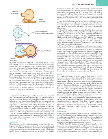
Fig. 149.6 CATALYSIS OF HEPARIN COFACTOR II (HCII) BY GLY- genital or acquired antithrombin deficiency and those with unusually
COSAMINOGLYCANS. In its unactivated form, the acidic N-terminal tail high levels of heparin-binding proteins often require very high doses
of HCII is tethered to the positively charged glycosaminoglycan-binding site of heparin to achieve a therapeutic aPTT or anti–factor Xa level. If
on the body of the serpin. The binding of heparin or dermatan sulfate to this there is good correlation between the aPTT and the anti–factor Xa
binding site displaces the N-terminal tail, thereby facilitating the interaction levels, either test can be used to monitor heparin therapy.
of this anionic domain with exosite 1 on thrombin. Glycosaminoglycan
binding also evokes a conformational change in the reactive center loop of Dosing
HCII that contributes to the formation of the HCII–thrombin complex. For prophylaxis, heparin is usually given in fixed doses of 5000 U
Longer heparin chains bind not only to HCII but also to the heparin-binding subcutaneously two or three times daily. With these low doses, coagu-
domain on thrombin, so-called exosite 2. Heparin-mediated bridging of lation monitoring is unnecessary. In contrast, monitoring is essential
HCII to thrombin enhances the rate of inhibition. Consequently, longer when heparin is given in therapeutic doses because a subtherapeutic
heparin chains that contain at least 26 saccharide units promote thrombin anticoagulant response has been associated with a higher risk of
inhibition by HCII to a greater extent than shorter chains. Unlike heparin, recurrent thrombosis. Fixed-dose or weight-based heparin nomo-
dermatan sulfate does not need to bind to thrombin for maximal enhance- grams are used to standardize heparin dosing and to shorten the time
ment in the rate of thrombin inhibition, because shorter dermatan sulfate required to achieve a therapeutic anticoagulant response. At least two
fragments are just as active as longer ones.
heparin nomograms have been validated in patients with venous
thromboembolism and reduce the time required to achieve a thera-
peutic aPTT. Weight-adjusted heparin nomograms also have been
Heparin is cleared through a combination of a rapid, saturable evaluated in patients with acute coronary syndromes.
and a much slower first-order mechanism. The saturable phase of Unmonitored twice-daily subcutaneous heparin proved as effec-
heparin clearance is thought to be caused by binding to endothelial tive and safe as once-daily LMWH for initial treatment of venous
15a
cell receptors and macrophages. Bound heparin is internalized and thromboembolism in one study. High doses of heparin were used;
depolymerized. The slower, nonsaturable mechanism of clearance is treatment was started with a dose of 333 U/kg followed by 250 U/
largely renal. At therapeutic doses, a large proportion of heparin is kg twice daily thereafter. This regimen may be an option for patients
cleared through the rapid, saturable, dose-dependent mechanism. with renal insufficiency where LMWH or fondaparinux is
The complex kinetics of clearance renders the anticoagulant response problematic.
to heparin nonlinear at therapeutic doses, with both the intensity and
duration of effect rising disproportionately with increasing dose. Limitations
Thus the apparent biological half-life of heparin increases from Heparin has pharmacokinetic and biophysical limitations (Table
approximately 30 minutes after an intravenous bolus of 25 U/kg, to 149.2). The pharmacokinetic limitations reflect heparin’s propensity
60 minutes with an intravenous bolus of 100 U/kg, to 150 minutes to bind in a pentasaccharide-independent fashion to cells and plasma
with a bolus of 400 U/kg. proteins. Heparin binding to cells explains its dose-dependent clear-
ance, whereas binding to plasma proteins results in a variable antico-
Monitoring agulant response and can lead to heparin resistance.
Heparin therapy can be monitored using the activated partial throm- The biophysical limitations of heparin reflect the inability of the
boplastin time (aPTT), anti–factor Xa level, or the activated clotting heparin–antithrombin complex (1) to inhibit factor Xa when it is
time (ACT). Although the aPTT is the test most often employed for incorporated into the prothrombinase complex, the complex that
this purpose, there are problems with this assay. aPTT reagents vary converts prothrombin to thrombin, and (2) to inhibit thrombin
in their sensitivity to heparin, and the type of coagulometer used for bound to fibrin. Consequently, factor Xa bound to activated platelets