
Page 250 - Hematology_ Basic Principles and Practice ( PDFDrive )
P. 250
202 Part III Immunologic Basis of Hematology
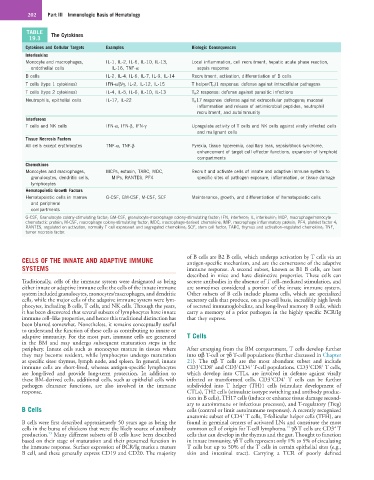
TABLE The Cytokines
19.3
Cytokines and Cellular Targets Examples Biologic Consequences
Interleukins
Monocyte and macrophages, IL-1, IL-2, IL-6, IL-10, IL-13, Local inflammation, cell recruitment, hepatic acute phase reaction,
endothelial cells IL-16, TNF-α sepsis response
B cells IL-2, IL-4, IL-6, IL-7, IL-9, IL-14 Recruitment, activation, differentiation of B cells
T cells (type 1 cytokines) IFN-α/β/γ, IL-2, IL-12, IL-15 T helper(T H )1 response: defense against intracellular pathogens
T cells (type 2 cytokines) IL-4, IL-5, IL-6, IL-10, IL-13 T H 2 response: defense against parasitic infections
Neutrophils, epithelial cells IL-17, IL-22 T H 17 response: defense against extracellular pathogens; mucosal
inflammation and release of antimicrobial peptides, neutrophil
recruitment, and autoimmunity
Interferons
T cells and NK cells IFN-α, IFN-β, IFN-γ Upregulate activity of T cells and NK cells against virally infected cells
and malignant cells
Tissue Necrosis Factors
All cells except erythrocytes TNF-α, TNF-β Pyrexia, tissue hyperemia, capillary leak, sepsis/shock syndrome,
enhancement of target cell effector functions, expansion of lymphoid
compartments
Chemokines
Monocytes and macrophages, MCPs, eotaxin, TARC, MDC, Recruit and activate cells of innate and adaptive immune system to
granulocytes, dendritic cells, MIPs, RANTES, PF4 specific sites of pathogen exposure, inflammation, or tissue damage
lymphocytes
Hematopoietic Growth Factors
Hematopoietic cells in marrow G-CSF, GM-CSF, M-CSF, SCF Maintenance, growth, and differentiation of hematopoietic cells
and peripheral
compartments
G-CSF, Granulocyte colony-stimulating factor; GM-CSF, granulocyte-macrophage colony-stimulating factor; IFN, interferon; IL, interleukin; MCP, macrophage/monocyte
chemotactic protein; M-CSF, macrophage colony-stimulating factor; MDC, macrophage-derived chemokine; MIP, macrophage inflammatory protein; PF4, platelet factor 4;
RANTES, regulated on activation, normally T cell expressed and segregated chemokine; SCF, stem cell factor; TARC, thymus and activation–regulated chemokine; TNF,
tumor necrosis factor.
CELLS OF THE INNATE AND ADAPTIVE IMMUNE of B cells are B2 B cells, which undergo activation by T cells via an
antigen-specific mechanism, and are the cornerstone of the adaptive
SYSTEMS immune response. A second subset, known as B1 B cells, are best
described in mice and have distinctive properties. These cells can
Traditionally, cells of the immune system were designated as being secrete antibodies in the absence of T cell–mediated stimulation, and
either innate or adaptive immune cells: the cells of the innate immune are sometimes considered a portion of the innate immune system.
system included granulocytes, monocytes/macrophages, and dendritic Other subsets of B cells include plasma cells, which are specialized
cells, while the major cells of the adaptive immune system were lym- secretory cells that produce, on a per-cell basis, incredibly high levels
phocytes, including B cells, T cells, and NK cells. Through the years, of secreted immunoglobulins, and long-lived memory B cells, which
it has been discovered that several subsets of lymphocytes have innate carry a memory of a prior pathogen in the highly specific BCR/Ig
immune cell–like properties, and hence this traditional distinction has that they express.
been blurred somewhat. Nonetheless, it remains conceptually useful
to understand the function of these cells as contributing to innate or
adaptive immunity. For the most part, immune cells are generated T Cells
in the BM and may undergo subsequent maturation steps in the
periphery. Innate cells such as monocytes mature in tissues where After emerging from the BM compartment, T cells develop further
they may become resident, while lymphocytes undergo maturation into αβ T-cell or γδ T-cell populations (further discussed in Chapter
at specific sites: thymus, lymph node, and spleen. In general, innate 21). The αβ T cells are the most abundant subset and include
+
+
+
+
+
+
immune cells are short-lived, whereas antigen-specific lymphocytes CD3 CD8 and CD3 CD4 T-cell populations. CD3 CD8 T cells,
are long-lived and provide long-term protection. In addition to which develop into CTLs, are involved in defense against virally
+
+
these BM-derived cells, additional cells, such as epithelial cells with infected or transformed cells. CD3 CD4 T cells can be further
pathogen clearance functions, are also involved in the immune subdivided into T helper (TH)1 cells (stimulate development of
response. CTLs), TH2 cells (stimulate isotype switching and antibody produc-
tion in B cells), TH17 cells (induce or enhance tissue damage second-
ary to autoimmune or infectious processes), and T-regulatory (Treg)
B Cells cells (control or limit autoimmune responses). A recently recognized
+
anatomic subset of CD4 T cells, T-follicular helper cells (TFH), are
B cells were first described approximately 50 years ago as being the found in germinal centers of activated LNs and constitute the most
+
14
cells in the bursa of chickens that were the likely source of antibody common cell of origin for T-cell lymphoma. γδ T cells are CD3 T
13
production. Many different subsets of B cells have been described cells that can develop in the thymus and the gut. Thought to function
based on their stage of maturation and their presumed function in in innate immunity, γδ T cells represent only 1% to 5% of circulating
the immune response. Surface expression of BCR/Ig marks a mature T cells but up to 50% of the T cells in certain epithelial sites (e.g.,
B cell, and these generally express CD19 and CD20. The majority skin and intestinal tract). Carrying a TCR of poorly defined