
Page 2540 - Hematology_ Basic Principles and Practice ( PDFDrive )
P. 2540
Chapter 157 Hematologic Manifestations of HIV/AIDS 2263
TABLE Surveillance Case Definition for HIV Infection in gp 41
157.1 Adults and Adolescents (Age >13 Years) gp 120
Stage Laboratory Evidence Clinical Evidence Lipid membrane
Stage1 Laboratory confirmation of No AIDS-defining
HIV infection and CD4 + condition (see Table Viral RNA p17
T lymphocyte count of 157.2)
+
≥500 cells/µL or CD4
T-lymphocyte p9
percentage of ≥29% a p7
Stage 2 Laboratory confirmation of No AIDS-defining
HIV infection and CD4 + condition (see Table p24
T lymphocyte count of 157.2)
200–499 cells/µL or
+
CD4 T-lymphocyte Reverse
percentage of 14–28% a transcriptase
Stage 3 Laboratory confirmation of Documentation of an
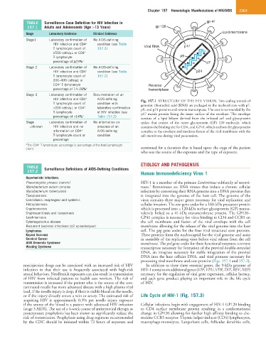
HIV infection and CD4 + AIDS-defining Fig. 157.1 STRUCTURE OF THE HIV VIRION. Two coding strands of
T lymphocyte count of condition with genomic ribonucleic acid (RNA) are packaged in the nucleoid core with p7,
+
<200 cells/µL or CD4 laboratory confirmation p9, and p24 proteins and reverse transcriptase. The core is surrounded by the
T-lymphocyte of HIV infection (see p17 matrix protein lining the inner surface of the envelope. The envelope
percentage of <14% a Table 157.2)
consists of a lipid bilayer derived from the infected cell and glycoprotein
Stage Laboratory confirmation of No information on spikes that consist of the outer glycoprotein (GP) 120 molecule, which
unknown HIV infection and no presence of an contains the binding site for CD4, and GP41, which anchors the glycoprotein
+
information on CD4 AIDS-defining complex to the envelope and mediates fusion of the viral membrane with the
T-lymphocyte count or condition cell membrane during viral penetration.
percentage
a The CD4 T-lymphocyte percentage is percentage of the total lymphocyte
+
count. continued for a duration that is based upon the stage of the patient
who was the source of the exposure and the type of exposure.
ETIOLOGY AND PATHOGENSIS
TABLE Surveillance Definitions of AIDS-Defining Conditions
157.2
Human Immunodeficiency Virus 1
Opportunistic Infections:
Pneumocystis jirovecii (carinii) HIV-1 is a member of the primate Lentivirinae subfamily of retrovi-
3
Mycobacterium avium complex ruses. Retroviruses are RNA viruses that induce a chronic cellular
Mycobacterium tuberculosis infection by converting their RNA genome into a DNA provirus that
Toxoplasmosis is integrated into the genome of the host cell. The genome of the
Candidiasis: esophageal and systemic virus contains three major genes necessary for viral replication and
Histoplasmosis cellular invasion. The env gene codes for a 160-kDa precursor protein
Cryptococcosis which is processed into a 120-kDa surface glycoprotein (GP) nonco-
Cryptosporidiosis and isosporiasis valently linked to a 41-kDa transmembrane protein. The GP120–
Leishmaniasis GP41 complex is necessary for virus binding to CD4 and CCR5 on
Cytomegalovirus disease the cell membrane and fusion of the viral envelope with the cell
Recurrent bacterial infections (≥2 episodes/year) membrane allowing for the release of the viral genome into the host
Lymphomas cell. The gag gene codes for the four viral structural core proteins.
Kaposi Sarcoma These proteins form the nucleocapsid for the viral genome and assist
Cervical Cancer in assembly of the replicating virus before viral release from the cell
AIDS Dementia Syndrome membrane. The pol gene codes for three functional enzymes; a reverse
Wasting Syndrome transcriptase necessary for formation of the proviral double-stranded
DNA, an integrase necessary for stable integration of the proviral
DNA into the host cellular DNA, and viral protease necessary for
processing viral membrane and core proteins (Figs. 157.1 and 157.2).
noninjection drugs can be associated with an increased risk of HIV In addition to these three essential genes, the 9-kDa genome of
infection in that their use is frequently associated with high-risk HIV-1 contains six additional genes (VIF, VPU, VPR, TAT, REV, NEF)
sexual behaviors. Needlestick exposures can also result in transmission necessary for the regulation of viral gene expression, cellular latency,
of HIV from infected patients to health care workers. The risk of and each gene product playing an important role in the life cycle
transmission is increased if the patient who is the source of the con- of HIV.
taminated needle has more advanced disease with a high plasma viral
load, if the needle injury is deep, if there is visible blood on the needle,
or if the injury directly enters a vein or artery. The estimated risk of Life Cycle of HIV-1 (Fig. 157.3)
acquiring HIV is approximately 0.3% per needle injury exposure
if the source of the blood is a patient with advanced HIV infection Cellular infections begin with engagement of HIV-1 GP120 binding
(stage 3 AIDS). The use of a 4-week course of antiretroviral therapy as to CD4 surface membrane protein resulting in a conformational
postexposure prophylaxis has been shown to significantly reduce the change in GP120 allowing for further high affinity binding to che-
risk of transmission. Prophylaxis using drug regimens recommended mokine CCR5 receptor. Thymic helper-inducer (CD4) lymphocytes,
by the CDC should be initiated within 72 hours of exposure and macrophage-monocytes, Langerhans cells, follicular dendritic cells,