
Page 2544 - Hematology_ Basic Principles and Practice ( PDFDrive )
P. 2544
Chapter 157 Hematologic Manifestations of HIV/AIDS 2267
the interval immediately following the start of therapy, there is a The bone marrow in patients with HIV can be hypercellular,
+
+
prompt increase in both CD4 and CD8 cells that is composed normocellular, or hypocellular. In a majority of cases the normal bone
+
predominantly of cells of a memory phenotype (CD45RO or marrow architecture is often disturbed, with dysplastic changes
+
+
–
6
CD45RA CD62L ). This increase is slightly different for CD4 cells, similar to those seen in MDS (Fig. 157.4). In Fig. 157.5 are shown
which increase more briskly (0.027/day) and plateau at approximately additional features seen in the bone marrow of HIV-infected indi-
+
3 weeks compared with CD8 cells (increase of 0.008/day), which viduals. HIV-associated stromal changes include edema, gelatinous
5
plateau at 8 weeks. This increase is thought to be largely caused by transformation, and increased reticulin fibers. Dense collagen fibrosis,
redistribution from peripheral tissues, perhaps related to a changing however, is not a feature of the HIV bone marrow. There are some
level of activation of the cells with declining viral antigen stimulation. important features distinguishing the morphology of the bone
This initial increase in circulating cell numbers does not achieve marrow of an HIV-infected individual from that of patients with
normal blood levels of lymphocytes. The secondary, much slower MDS. Dyserythropoiesis is usually less severe in HIV than observed
phase of T-cell increase tends to be sustained for months to years, in MDS, and occurs predominantly in patients treated with HAART.
+
with a greater contribution of cells with a naive phenotype (CD45RA Megaloblastic changes are usually associated with zidovudine and
+
CD62L ). The naive population rises along with cells bearing the stavudine therapy. In contrast to MDS, in which erythropoiesis may
T-cell receptor excision circle, an indicator of recent T-cell receptor be hyperplastic, the myeloid/erythroid ratio in HIV is usually normal
rearrangement that accompanies early T-cell differentiation. It is this and there may even be neutrophilic and megakaryocytic hyperplasia.
population that is generally regarded as thymus dependent and that
is capable of truly expanding the immune repertoire. In addition, in
vivo models have further defined that T-cell generation, from precur-
sor populations both endogenous and exogenous to the thymus, BOX 157.2 Peripheral Blood Smear and Bone Marrow Morphology in
accompanies control of viremia. HIV/AIDS
The peripheral blood smear of a patient with HIV/AIDS might show
Evaluation of Cytopenias in HIV-Infected Individuals anisocytosis, poikilocytosis, and rouleaux formation. Anemia, when
present, is usually normocytic and normochromic. Sometimes macro-
cytic can be seen even in the absence of AZT therapy. Lymphopenia
The evaluation of cytopenias in patients infected with HIV requires is seen in advanced disease. Hypogranular neutrophils and Pelger
a review of complete blood count and thorough examination of the forms are rarely present. Platelets can be normal or hypogranular. In
peripheral blood smear (see box on Peripheral Blood Smear and cases of thrombocytopenia, the platelets can be normal sized or large
Bone Marrow Morphology in HIV/AIDS). Although there is a when thrombocytopenia is caused by immune destruction with perse-
+
gradual fall in CD4 lymphocytes during the asymptomatic phase vered marrow.
of HIV infection, a mild lymphocytosis may at times be seen, The bone marrow is usually hypercellular, but can be normocellular
+
caused by an increase in CD8 lymphocytes. Atypical or activated or hypocellular. Interstitial and perivascular polyclonal plasmacytosis is
lymphocytes may frequently be seen. Lymphopenia is present in the usually present. HIV-associated stromal changes include edema,
advanced stage of the disease. Anemia, when present, is usually gelatinous transformation and increased reticulin fibers (dense collagen
fibers are not a feature of HIV). Normal bone marrow architecture is
normocytic and normochromic. It can be at times macrocytic, often disturbed and dysplastic changes can be seen, including dys-
either because of the effect of certain antiretroviral drugs such as erythropoiesis, dysgranulopoiesis, and abnormal megakaryocytes
zidovudine or stavudine, or seen in patients with advanced HIV (including clustures and bare megakaryocytic nuclei). However, the
disease. Occasionally red blood cell (RBC) anisocytosis, poikilocyto- following features distinguish the bone marrow morphology in HIV from
sis, and rouleaux can sometimes be seen in patients with untreated that of MDS: dysplasia is less severe in HIV. Dyserythropoiesis occurs
advanced HIV disease. Also hypogranular neutrophils and Pelger- mainly in patients on HAART. Megaloblastic changes are associated
Huët forms may rarely be present in patients with advanced HIV with AZT therapy. Although erythropoiesis is usually hyperplastic in
disease. However, in comparison to patients with myelodysplasia MDS, myeloid to erythroid ratio is usually normal in HIV. Increase blasts
(MDS), agranular neutrophils and neutrophils with the acquired can be seen in MDS but never in HIV. Lastly, in contrast to MDS, the
bone marrow in HIV often shows eosinophilia, lymphohistiocytic infil-
Pelger-Huët anomaly are not a predominant feature in patients with trates and plasmacytosis.
HIV infection. Thrombocytopenia is associated with normal sized
platelets, except occasional large platelets may be seen when there is AIDS, Acquired immunodeficiency syndrome; AZT, zidovudine; HAART, highly
immune-mediated destruction of platelets with preserved marrow active antiretroviral therapy; HIV, human immunodeficiency virus; MDS,
function. myelodysplastic syndrome.
A B C D D
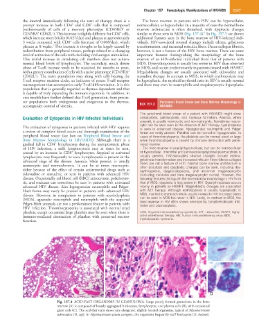
Fig. 157.4 ACID-FAST ORGANISMS IN GRANULOMA. Large poorly formed granuloma in the bone
marrow (A) is composed of loosely aggregated histiocytes, lymphocytes, and plasma cells (B), with occasional
giant cells (C). The acid-fast stain shows rare elongated, slightly beaded organisms, typical of Mycobacterium
tuberculosis (D, top). In Mycobacterium avium complex, the organisms frequently stuff histiocytes (D, bottom).