
Page 414 - Hematology_ Basic Principles and Practice ( PDFDrive )
P. 414
Chapter 28 Thrombocytopoiesis 335
Mature Megakaryocytes
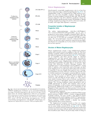
CFC-Mk-HPP (?) Morphologically recognizable megakaryocytes exist in at least four
distinct maturation stages as defined morphologically (Fig. 28.2). The
megakaryoblast (stage I) is characterized by a high nucleus-to-cyto-
plasm ratio and scanty basophilic cytoplasm, reflecting the large
Proliferating BFU-Mk amount of protein synthesis occurring in these cells. The promega-
megakaryocytes karyocyte (stage II) is the cell in which the cytoplasmic volume and
(2N/4N) number of platelet-specific granules increase. The granular or “platelet
shedding” megakaryocyte (stages III and IV) is the most mature cell.
In reality, these stages likely represent a continuum.
CFU-Mk
Prospective Isolation of Megakaryocyte
Progenitor Cells
PMkB The surface immunophenotype: c-kit(+)Sca-1(-)IL7Ralpha(-)
Thy1.1(-)Lin(-)CD9(+)CD41(+)FcgammaR(lo) can be used to
prospectively isolate murine clonogenic committed MkPs. This frac-
tion represents approximately 0.01% of the total nucleated BM cells
Immature and gives rise to CFU-Mk and occasionally BFU-Mk in colony
(transitional) assays. The immunophenotype Lin(-)c-kit (+)Sca1(-)CD150 (+)
megakaryocytes PMkB CD41(+) has also been used to enrich for committed murine MkPs.
(4N-8N)
Identification of a comparable set of surface markers for human MkPs
has not been reported.
PMkB
Structure of Mature Megakaryocytes
Mature megakaryocytes contain a large multilobulated polyploid
nucleus often situated toward the periphery of the cell. They have
abundant cytoplasm, which contains platelet-specific secretory gran-
Stage I 2
ules, alpha (α-) granules and dense granules (Fig. 28.3). The biogen-
esis of α-granules and dense granules begins in immature
megakaryocytes, and both granule types develop concomitantly.
α-Granules are 200 to 500 nm in diameter and have a dense center
and fine granular matrix. Megakaryocytes synthesize many of the
Mature, postmitotic constituents of α-granules and target them to the granules. These
megakaryocytes Stage II include vWF, fibronectin, P-selectin, fibrinogen receptors, PF4,
(8N-128N) coagulation factor V, and plasminogen activator inhibitor-1, among
others. In addition, some constituents, such as fibrinogen, are taken
up by megakaryocytes via endocytosis and/or pinocytosis and stored
in α-granules. It was once thought that α-granules were a homoge-
neous population of vesicles. However, it has become clear that there
are distinct populations of α-granules containing different constitu-
Stage III/IV ents, and that these can be differentially released during platelet
3
activation. Dense granules are 200 to 300 nm in diameter and
consist of a halo encircling an electron opaque core. They contain
many soluble hemostatic factors such as serotonin, catecholamines,
adenosine, adenosine 5′-diphosphate, adenosine 5′-triphosphate, and
calcium. Their limiting membranes contain glycoproteins such as
αIIbβ3, glycoprotein Ib (GPIb), and P-selectin, which are also
Platelets present in α-granules, as well as unique membrane proteins such as
granulophysin. Multivesicular bodies serve as intermediates in the
biogenesis of both α-granules and dense granules. It has been pro-
Fig. 28.1 CELLULAR HIERARCHY OF MEGAKARYOCYTE DEVEL- posed that they constitute a sorting compartment between α-granule
OPMENT. Megakaryocyte development can be conceptually divided into and dense granule components.
three stages: The proliferating progenitor cells, which have the typical 2N/4N Mutations in the NBEAL2 gene have recently been linked to gray
DNA content; the immature megakaryocytes, which have an intermediate platelet syndrome (OMIM 139090), a disorder of impaired platelet
DNA content and are transitional between the progenitor cells and the more α-granule synthesis. This gene encodes a large BEACH domain
mature cells; and the mature, postmitotic cells, which have an 8N to 128N containing protein that shares homology with the LYST gene product.
DNA content. BFU-Mk, Burst-forming unit-megakaryocyte; CFU-Mk, LYST is involved in vesicular trafficking and is mutated in Chediak-
colony-forming unit-megakaryocyte; CFC-Mk-HPP, colony-forming unit- Higashi syndrome (OMIM 214500), a disorder that includes
megakaryocyte high-proliferative potential; PMkB, promegakaryoblast. impaired platelet dense granule biogenesis.
The megakaryocyte cytoplasm contains at least two complex
membranous systems: the demarcation membrane system (DMS)
and the dense tubular network (DTS) (see Fig. 28.3). The DMS
consists of an extensive network of tubular and flattened membranous
structures that interconnect with one another and communicate with
the extracellular space. Whole cell patch-clamp studies in living rat