
Page 591 - Hematology_ Basic Principles and Practice ( PDFDrive )
P. 591
506 Part V Red Blood Cells
A B
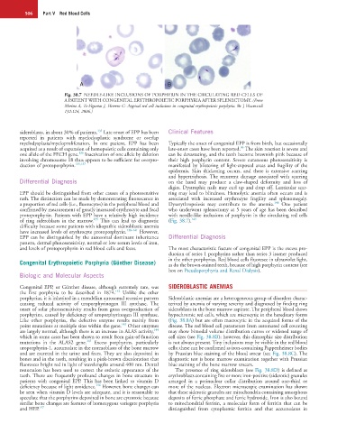
Fig. 38.7 NEEDLE-LIKE INCLUSIONS OF PORPHYRIN IN THE CIRCULATING RED CELLS OF
A PATIENT WITH CONGENITAL ERYTHROPOIETIC PORPHYRIA AFTER SPLENECTOMY. (From
Merino A, To-Figueras J, Herrero C: Atypical red cell inclusions in congenital erythropoietic porphyria. Br J Haematol
132:124, 2006.)
135
sideroblasts, in about 30% of patients. Late onset of EPP has been Clinical Features
reported in patients with myelodysplastic syndrome or overlap
myelodysplasia/myeloproliferation. In one patient, EPP has been Typically the onset of congenital EPP is from birth, but occasionally
96
acquired as a result of expansion of hemopoietic cells containing only late-onset cases have been reported. The skin reaction is severe and
136
one allele of the FECH gene. Inactivation of one allele by deletion can be devastating, and the teeth become brownish pink because of
involving chromosome 18 thus appears to be sufficient for overpro- their high porphyrin content. Severe cutaneous photosensitivity is
duction of protoporphyrin. 135,137 manifested by blistering of light-exposed areas and fragility of the
epidermis. Skin thickening occurs, and there is extensive scarring
and hypertrichosis. The recurrent damage associated with scarring
Differential Diagnosis on the hand may produce a claw-shaped deformity and loss of
digits. Dystrophic nails may curl up and drop off. Lenticular scar-
EPP should be distinguished from other causes of a photosensitive ring may lead to blindness. Hemolytic anemia often occurs and is
rash. The distinction can be made by demonstrating fluorescence in associated with increased erythrocyte fragility and splenomegaly.
148
a proportion of red cells (i.e., fluorocytes) in the peripheral blood and Dyserythropoiesis may contribute to the anemia. One patient
confirmed by measurement of greatly increased erythrocyte and fecal who underwent splenectomy at 5 years of age has been described
protoporphyrin. Patients with EPP have a relatively high incidence with needle-like inclusions of porphyrin in the circulating red cells
135
of ring sideroblasts in the marrow. This can lead to diagnostic (Fig. 38.7). 149
difficulty because some patients with idiopathic sideroblastic anemia
have increased levels of erythrocyte protoporphyrin. 138–141 However,
EPP can be distinguished by the autosomal dominant inheritance Differential Diagnosis
pattern, dermal photosensitivity, normal or low serum levels of iron,
and levels of protoporphyrin in red blood cells and feces. The most characteristic feature of congenital EPP is the excess pro-
duction of series 1 porphyrins rather than series 3 isomer produced
Congenital Erythropoietic Porphyria (Günther Disease) in the other porphyrias. Red blood cells fluoresce in ultraviolet light,
as do the brown-stained teeth, because of high porphyrin content (see
box on Pseudoporphyria and Renal Dialysis).
Biologic and Molecular Aspects
Congenital EPP, or Günther disease, although extremely rare, was SIDEROBLASTIC ANEMIAS
142
the first porphyria to be described in 1874. Unlike the other
porphyrias, it is inherited in a mendelian autosomal recessive pattern Sideroblastic anemias are a heterogeneous group of disorders charac-
causing reduced activity of uroporphyrinogen III synthase. The terized by anemia of varying severity and diagnosed by finding ring
onset of solar photosensitivity results from gross overproduction of sideroblasts in the bone marrow aspirate. The peripheral blood shows
porphyrins, caused by deficiency of uroporphyrinogen III synthase. hypochromic red cells, which are microcytic in the hereditary forms
Like other porphyrias, the defective enzyme results mainly from (Fig. 38.8A) but are often macrocytic in the acquired forms of the
143
point mutations at multiple sites within the gene. Other enzymes disease. The red blood cell parameters from automated cell counting
144
are largely normal, although there is an increase in ALAS activity, may show bimodal volume distribution curves or widened range of
which in some cases has been shown to result from gain-of-function cell sizes (see Fig. 38.8B); however, this dimorphic size distribution
145
mutations in the ALAS2 gene. Excess porphyrins, particularly is not always present. Tiny inclusions may be visible in the red blood
uroporphyrin-1, accumulate in the normoblasts of the bone marrow cells; these can be confirmed as iron-containing Pappenheimer bodies
and are excreted in the urine and feces. They are also deposited in by Prussian blue staining of the blood smear (see Fig. 38.8C). The
bones and in the teeth, resulting in a pink-brown discoloration that diagnostic test is bone marrow examination together with Prussian
fluoresces bright red in light of wavelengths around 400 nm. Dental blue staining of the bone marrow smears.
restoration has been used to correct the esthetic appearance of the The presence of ring sideroblasts (see Fig. 38.8D) is defined as
teeth. There are frequently profound changes in bone structure in erythroblasts containing five or more iron-positive (siderotic) granules
patients with congenital EPP. This has been linked to vitamin D arranged in a perinuclear collar distribution around one-third or
146
deficiency because of light avoidance. However, bone changes can more of the nucleus. Electron microscopic examination has shown
be seen when vitamin D levels are adequate, and it is reasonable to that these siderotic granules are mitochondria containing amorphous
speculate that the porphyrins deposited in bone are cytotoxic because deposits of ferric phosphate and ferric hydroxide. Iron is also bound
similar bone changes are features of homozygous variegate porphyria to mitochondrial ferritin, a molecular form of ferritin that can be
and HEP. 147 distinguished from cytoplasmic ferritin and that accumulates in