
Page 849 - Hematology_ Basic Principles and Practice ( PDFDrive )
P. 849
732 Part VI Non-Malignant Leukocytes
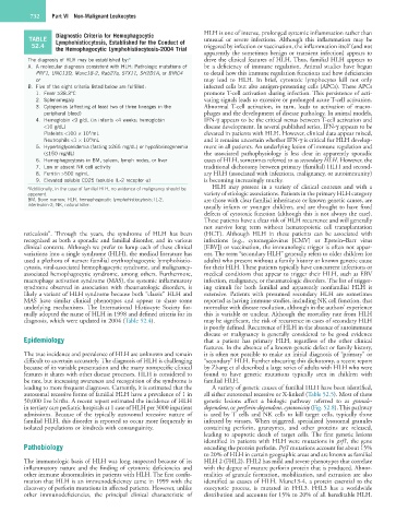
Diagnostic Criteria for Hemophagocytic HLH is one of intense, prolonged systemic inflammation rather than
TABLE Lymphohistiocytosis, Established for the Conduct of unusual or severe infections. Although this inflammation may be
52.4 the Hemophagocytic Lymphohistiocytosis-2004 Trial triggered by infection or vaccination, the inflammation itself (and not
apparently the sometimes benign or transient infection) appears to
The diagnosis of HLH may be established by: a drive the clinical features of HLH. Thus, familial HLH appears to
A. A molecular diagnosis consistent with HLH: Pathologic mutations of be a deficiency of immune regulation. Animal studies have begun
PRF1, UNC13D, Munc18-2, Rab27a, STX11, SH2D1A, or BIRC4 to detail how this immune regulation functions and how deficiencies
or may lead to HLH. In brief, cytotoxic lymphocytes kill not only
B. Five of the eight criteria listed below are fulfilled: infected cells but also antigen-presenting cells (APCs). These APCs
1. Fever ≥38.3°C promote T-cell activation during infection. This persistence of acti-
2. Splenomegaly vating signals leads to excessive or prolonged acute T-cell activation.
3. Cytopenias (affecting at least two of three lineages in the Abnormal T-cell activation, in turn, leads to activation of macro-
peripheral blood) phages and the development of disease pathology. In animal models,
4. Hemoglobin <9 g/dL (in infants <4 weeks: hemoglobin IFN-γ appears to be the critical nexus between T-cell activation and
<10 g/dL) disease development. In several published series, IFN-γ appears to be
Platelets <100 × 10 /mL elevated in patients with HLH. However, clinical data appear mixed,
3
Neutrophils <1 × 10 /mL and it remains uncertain whether IFN-γ is critical for HLH develop-
3
5. Hypertriglyceridemia (fasting ≥265 mg/dL) or hypofibrinogenemia ment in all patients. An underlying lesion of immune regulation and
(≤150 mg/dL) the associated pathophysiology is less clear in apparently sporadic
6. Hemophagocytosis in BM, spleen, lymph nodes, or liver cases of HLH, sometimes referred to as secondary HLH. However, the
7. Low or absent NK cell activity traditional dichotomy between primary (familial) HLH and second-
8. Ferritin >500 ng/mL ary HLH (associated with infections, malignancy, or autoimmunity)
9. Elevated soluble CD25 (soluble IL-2 receptor α) is becoming increasingly murky.
a Additionally, in the case of familial HLH, no evidence of malignancy should be HLH may present in a variety of clinical contexts and with a
apparent. variety of etiologic associations. Patients in the primary HLH category
BM, Bone marrow; HLH, hemophagocytic lymphohistiocytosis; IL-2, are those with clear familial inheritance or known genetic causes, are
interleukin-2; NK, natural killer. usually infants or younger children, and are thought to have fixed
defects of cytotoxic function (although this is not always the case).
These patients have a clear risk of HLH recurrence and will generally
not survive long term without hematopoietic cell transplantation
reticulosis”. Through the years, the syndrome of HLH has been (HCT). Although HLH in these patients can be associated with
recognized as both a sporadic and familial disorder, and in various infections (e.g., cytomegalovirus [CMV] or Epstein–Barr virus
clinical contexts. Although we prefer to lump each of these clinical [EBV]) or vaccination, the immunologic trigger is often not appar-
variations into a single syndrome (HLH), the medical literature has ent. The term “secondary HLH” generally refers to older children (or
used a plethora of names: familial erythrophagocytic lymphohistio- adults) who present without a family history or known genetic cause
cytosis, viral-associated hemophagocytic syndrome, and malignancy- for their HLH. These patients typically have concurrent infections or
associated hemophagocytic syndrome, among others. Furthermore, medical conditions that appear to trigger their HLH, such as EBV
macrophage activation syndrome (MAS), the systemic inflammatory infection, malignancy, or rheumatologic disorders. The list of trigger-
syndrome observed in association with rheumatologic disorders, is ing stimuli for both familial and apparently nonfamilial HLH is
likely a variant of HLH syndrome because both “classic” HLH and extensive. Patients with presumed secondary HLH are sometimes
MAS have similar clinical phenotypes and appear to share some reported as having immune studies, including NK cell function, that
underlying mechanisms. The International Histiocyte Society for- normalize with disease resolution, although in the authors’ experience
mally adopted the name of HLH in 1998 and defined criteria for its this is variable or unclear. Although the mortality rate from HLH
diagnosis, which were updated in 2004 (Table 52.4). may be significant, the risk of recurrence in cases of secondary HLH
is poorly defined. Recurrence of HLH in the absence of autoimmune
disease or malignancy is generally considered to be good evidence
Epidemiology that a patient has primary HLH, regardless of the other clinical
features. In the absence of a known genetic defect or family history,
The true incidence and prevalence of HLH are unknown and remain it is often not possible to make an initial diagnosis of “primary” or
difficult to ascertain accurately. The diagnosis of HLH is challenging “secondary” HLH. Further obscuring this dichotomy, a recent report
because of its variable presentation and the many nonspecific clinical by Zhang et al described a large series of adults with HLH who were
features it shares with other disease processes. HLH is considered to found to have genetic mutations typically seen in children with
be rare, but increasing awareness and recognition of the syndrome is familial HLH.
leading to more frequent diagnoses. Currently, it is estimated that the A variety of genetic causes of familial HLH have been identified,
autosomal recessive forms of familial HLH have a prevalence of 1 in all either autosomal recessive or X-linked (Table 52.5). Most of these
50,000 live births. A recent report estimated the incidence of HLH genetic lesions affect a biologic pathway referred to as granule-
in tertiary care pediatric hospitals at 1 case of HLH per 3000 inpatient dependent, or perforin-dependent, cytotoxicity (Fig. 52.8). This pathway
admissions. Because of the typically autosomal recessive nature of is used by T cells and NK cells to kill target cells, typically those
familial HLH, this disorder is reported to occur more frequently in infected by viruses. When triggered, specialized lysosomal granules
isolated populations or kindreds with consanguinity. containing perforin, granzymes, and other proteins are released,
leading to apoptotic death of target cells. The first genetic lesions
identified in patients with HLH were mutations in prf1, the gene
Pathobiology encoding the protein perforin. Prf1 mutations account for about 15%
to 20% of HLH in certain geographic areas and are known as familial
The immunologic basis of HLH was long suspected because of its HLH 2 (FHL2). FHL2 has mild and severe phenotypes that correlate
inflammatory nature and the finding of cytotoxic deficiencies and with the degree of mature perforin protein that is produced. Abnor-
other immune abnormalities in patients with HLH. The first confir- malities of granule formation, mobilization, and extrusion are also
mation that HLH is an immunodeficiency came in 1999 with the identified as causes of HLH. Munc13-4, a protein essential to the
discovery of perforin mutations in affected patients. However, unlike exocytotic process, is mutated in FHL3. FHL3 has a worldwide
other immunodeficiencies, the principal clinical characteristic of distribution and accounts for 15% to 20% of all hereditable HLH.