
Page 850 - Hematology_ Basic Principles and Practice ( PDFDrive )
P. 850
Chapter 52 Histiocytic Disorders 733
TABLE Hemophagocytic Lymphohistiocytosis-Associated Gene defects in SH2D1A or SAP. This disorder is a complex one character-
52.5 Mutations ized by lymphoproliferation, hypogammaglobulinemia, excess risk
of lymphoma, and development of HLH. It appears to share some
Gene Location Disease of the pathophysiology with classic familial HLH, although other
PRF1 10q21-22 FHL2 abnormalities (of B cells and other cells) make this disorder distinc-
tive. The second X-linked cause of HLH, sometimes called “XLP2”
UNC13D 17q25 FHL3
(although this is disputed), are abnormalities of a gene/protein called
STX11 6q24 FHL4 baculoviral inhibitor of apoptosis protein repeat-containing protein 4
RAB27A 15q21 Griscelli syndrome (BIRC4)/X-linked inhibitor of apoptosis protein (XIAP). The appar-
ently unique pathophysiology of HLH caused by XIAP deficiency
STXBP2 19p13 FHL5
is not understood. Other immunodeficiency syndromes caused by
Unknown 9q21.3-22 FHL1 defects in lysosomal trafficking have been linked to life-threatening
SH2D1A Xq24-26 XLP1 episodes of HLH. These include Chediak-Higashi syndrome, Gris-
XIAP Xq25 XLP2/X-linked HLH celli syndrome, and Hermansky-Pudlak syndrome, type II.
Taken together, the nine genetic disorders described above still
FHL, Familial hemophagocytic lymphohistiocytosis; HLH, hemophagocytic account for fewer than half of the diagnosed cases of HLH in children,
lymphohistiocytosis; XLP, X-linked lymphoproliferative syndrome.
including many familial cases still awaiting molecular definition.
Until recently, it was widely believed that symptoms of HLH trig-
gered by genetic causes arose during infancy and early childhood.
With the more widespread availability of genetic testing, it is apparent
Target cell
Apoptosis that the first significant episode of HLH can occur throughout life
from prenatal presentations through the seventh decade of life. Dis-
Granzyme B
Perforin tinctions between primary (genetically determined) and secondary
Cascade of
caspases (acquired) forms of HLH become increasingly blurred together as
new genetic causes are identified and patients who develop HLH
Effector cell
(CTL or NK cell) beyond early childhood or in the contexts of EBV infection or
Syntaxin 11 t-SNARE autoimmune disease are being found to share some of the same
Munc18-2
v-SNARE genetic etiologies.
Munc13-4 Clinical Manifestations
Rab27a Fusion
Priming
The classic presentation of HLH consists of prolonged, hectic fevers
Docking
(usually present for 1–2 weeks before diagnosis), hepatosplenomegaly,
and cytopenias. Neurologic symptoms are common and distinctive
Cytolytic features of these patients; these include symptoms of irritability,
granules ataxia, hypotonia or hypertonia, evidence of increased intracranial
pressure, meningismus, depressed mental status, cranial nerve palsies,
and seizures. Standard diagnostic criteria have been defined by the
Histiocyte Society for the conduct of the now closed HLH2004 trial
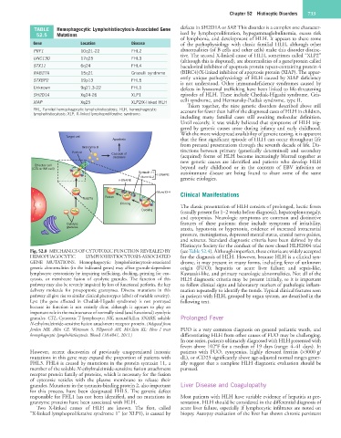
Fig. 52.8 MECHANICS OF CYTOTOXIC FUNCTION REVEALED BY (see Table 52.4). Although imperfect, these criteria are widely accepted
HEMOPHAGOCYTIC LYMPHOHISTIOCYTOSIS-ASSOCIATED for the diagnosis of HLH. However, because HLH is a clinical syn-
GENE MUTATIONS. Hemophagocytic lymphohistiocytosis-associated drome, it may present in many forms, including fever of unknown
genetic abnormalities (in the indicated genes) may affect granule-dependent origin (FUO); hepatitis or acute liver failure; and sepsis-like,
lymphocyte cytotoxicity by impairing trafficking, docking, priming for exo- Kawasaki-like, and primary neurologic abnormalities. Not all of the
cytosis, or membrane fusion of cytolytic granules. The function of this HLH diagnostic criteria may be present initially, so it is important
pathway may also be severely impaired by loss of functional perforin, the key to follow clinical signs and laboratory markers of pathologic inflam-
delivery molecule for proapoptotic granzymes. Diverse mutations in this mation repeatedly to identify the trends. Typical clinical features seen
pathway all give rise to similar clinical phenotypes (albeit of variable severity). in patients with HLH, grouped by organ system, are described in the
Lyst (the gene affected in Chediak-Higashi syndrome) is not portrayed following text.
because its function is not entirely clear, although it appears to play an
important role in the maintenance of normally sized (and functional) cytolytic
granules. CTL, Cytotoxic T lymphocyte; NK, natural killer, SNARE, soluble Prolonged Fever
N-ethylmaleimide-sensitive fusion attachment receptor protein. (Adapted from
Jordan MB, Allen CE, Weitzman S, Filipovich AH, McClain KL: How I treat FUO is a very common diagnosis on general pediatric wards, and
hemophagocytic lymphohistiocytosis. Blood 118:4041, 2011.) differentiating HLH from other causes of FUO may be challenging.
In one series, patients ultimately diagnosed with HLH presented with
fevers above 102°F for a median of 19 days (range: 4–41 days). In
However, recent discoveries of previously unappreciated intronic patients with FUO, cytopenias, highly elevated ferritin (>3000 g/
mutations in this gene may expand the proportion of patients with dL), or sCD25 significantly above age-adjusted normal ranges gener-
FHL3. FHL4 is caused by mutations in the protein syntaxin 11, a ally suggest that a complete HLH diagnostic evaluation should be
member of the soluble N-ethylmaleimide-sensitive fusion attachment pursued.
receptor protein family of proteins, which is necessary for the fusion
of cytotoxic vesicles with the plasma membrane to release their
granules. Mutations in the syntaxin-binding protein 2, also important Liver Disease and Coagulopathy
for this process, have been designated FHL5. The genetic defect
responsible for FHL1 has not been identified, and no mutations in Most patients with HLH have variable evidence of hepatitis at pre-
granzyme proteins have been associated with HLH. sentation. HLH should be considered in the differential diagnosis of
Two X-linked causes of HLH are known. The first, called acute liver failure, especially if lymphocytic infiltrates are noted on
“X-linked lymphoproliferative syndrome 1” (or XLP1), is caused by biopsy. Autopsy evaluation of the liver has shown chronic persistent