
Page 871 - Hematology_ Basic Principles and Practice ( PDFDrive )
P. 871
754 Part VI Non-Malignant Leukocytes
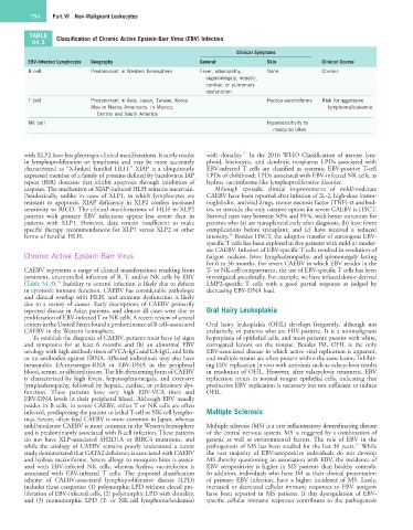
TABLE Classification of Chronic Active Epstein-Barr Virus (EBV) Infection
54.3
Clinical Symptoms
EBV-Infected Lymphocyte Geography General Skin Clinical Course
B cell Predominant in Western hemisphere Fever, adenopathy, None Chronic
organomegaly, hepatic,
cardiac, or pulmonary
dysfunction
T cell Predominant in Asia: Japan, Taiwan, Korea Hydroa vacciniforme Risk for aggressive
Also in Native Americans: in Mexico, lymphoma/leukemia
Central and South America
NK cell Hypersensitivity to
mosquito bites
17
with XLP2 have less pleotropic clinical manifestations. It rarely results with clonality. In the 2016 WHO Classification of mature lym-
in lymphoproliferation or lymphoma and may be more accurately phoid, histiocytic, and dendritic neoplasms LPDs associated with
characterized as “X-linked familial HLH.” XIAP is a ubiquitously EBV-infected T cells are classified as systemic EBV-positive T-cell
expressed member of a family of proteins defined by baculovirus IAP LPDs of childhood; LPDs associated with EBV-infected NK cells, as
repeat (BIR) domains that inhibit apoptosis through inhibition of hydroa vacciniforme-like lymphoproliferative disorder.
caspases. The mechanism of XIAP-induced HLH remains uncertain. Although sporadic clinical improvements of mild/moderate
Paradoxically, unlike in cases of XLP1, in which lymphocytes are CAEBV have been reported after infusion of IL-2, high-dose immu-
resistant to apoptosis, XIAP deficiency in XLP2 confers increased noglobulin, antiviral drugs, tumor necrosis factor (TNF)-α antibod-
sensitivity to RICD. The clinical manifestations of HLH in XLP2 ies, or steroids, the only curative option for severe CAEBV is HSCT.
patients with primary EBV infections appear less severe than in Survival rates vary between 50% and 95%, with better outcomes for
patients with XLP1. However, data remain insufficient to make patients who (a) are transplanted early after diagnosis, (b) have fewer
specific therapy recommendations for XLP1 versus XLP2 or other complications before transplant, and (c) have received a reduced
18
forms of familial HLH. intensity. Besides HSCT, the adoptive transfer of autologous EBV-
specific T cells has been explored in five patients with mild or moder-
ate CAEBV. Infusion of EBV-specific T cells resulted in resolution of
Chronic Active Epstein-Barr Virus fatigue, malaise, fever, lymphadenopathy, and splenomegaly lasting
for 6 to 36 months. For severe CAEBV in which EBV resides in the
CAEBV represents a range of clinical manifestations resulting from T- or NK-cell compartment, the use of EBV-specific T cells has been
persistent, uncontrolled infection of B, T, and/or NK cells by EBV investigated anecdotally. For example, we have infused donor-derived
16
(Table 54.3). Inability to control infection is likely due to defects LMP2-specific T cells with a good partial response as judged by
in cytotoxic immune function. CAEBV has considerable pathologic decreasing EBV-DNA load.
and clinical overlap with HLH, and immune dysfunction is likely
due to a variety of causes. Early descriptions of CAEBV primarily
reported disease in Asian patients, and almost all cases were due to Oral Hairy Leukoplakia
proliferation of EBV-infected T or NK cells. A recent review of several
centers in the United States found a predominance of B cell–associated Oral hairy leukoplakia (OHL) develops frequently, although not
CAEBV in the Western hemisphere. exclusively, in patients who are HIV-positive. It is a nonmalignant
To establish the diagnosis of CAEBV, patients must have (a) signs hyperplasia of epithelial cells, and most patients present with white,
and symptoms for at least 6 months and (b) an abnormal EBV corrugated lesions on the tongue. Besides IM, OHL is the only
serology with high antibody titers of VCA-IgG and EA-IgG, and little EBV-associated disease in which active viral replication is apparent,
or no antibodies against EBNA. Affected individuals may also have and multiple strains are often present within the same lesion. Inhibit-
measurable EA-messenger-RNA or EBV-DNA in the peripheral ing EBV replication in vivo with antivirals such as valacyclovir results
blood, serum, or affected tissues. The life-threatening form of CAEBV in resolution of OHL. However, after valacyclovir treatment, EBV
is characterized by high fevers, hepatosplenomegaly, and extensive replication recurs in normal tongue epithelial cells, indicating that
lymphadenopathy, followed by hepatic, cardiac, or pulmonary dys- productive EBV replication is necessary but not sufficient to induce
function. These patients have very high EBV-VCA titers and OHL.
EBV-DNA levels in their peripheral blood. Although EBV usually
resides in B cells, in severe CAEBV, either T or NK cells are often
infected, predisposing the patient to lethal T-cell or NK-cell lympho- Multiple Sclerosis
mas. Severe, often fatal CAEBV is more common in Japan, whereas
mild/moderate CAEBV is more common in the Western hemisphere Multiple sclerosis (MS) is a rare inflammatory demyelinating disease
and is predominantly associated with B-cell infection. These patients of the central nervous system. MS is triggered by a combination of
do not have XLP-associated SH2D1A or BIRC4 mutations, and genetic as well as environmental factors. The role of EBV in the
19
while the etiology of CAEBV remains poorly understood a recent pathogenesis of MS has been studied for the last 30 years. While
study demonstrated that GATA2 deficiency is associated with CAEBV the vast majority of EBV-seropositive individuals do not develop
and hydroa vacciniforme. Severe allergy to mosquito bites is associ- MS thereby questioning an association with EBV, the incidence of
ated with EBV-infected NK cells, whereas hydroa vacciniforme is EBV seropositivity is higher in MS patients than healthy controls.
associated with EBV-infected T cells. The proposed classification In addition, individuals who have IM as their clinical presentation
scheme of CAEBV-associated lymphoproliferative disease (LPD) of primary EBV infection, have a higher incidence of MS. Lastly,
includes three categories: (1) polymorphic LPD without clonal pro- increased or decreased cellular immune responses to EBV antigens
liferation of EBV-infected cells, (2) polymorphic LPD with clonality, have been reported in MS patients. If this dysregulation of EBV-
and (3) monomorphic LPD (T- or NK-cell lymphoma/leukemia) specific cellular immune responses contributes to the pathogenesis