
Page 929 - Hematology_ Basic Principles and Practice ( PDFDrive )
P. 929
812 Part VII Hematologic Malignancies
NPM1/MLF1 chimeric gene, which includes approximately half of
the 24-kb NPM1 gene, localized on 5q35, extending from exons 1–6,
juxtaposed to virtually the entire 35-kb MLF1 gene, localized on
3q25, starting at exon 2. The chimeric NPM1/MLF1 fusion protein
totals 426 amino acids with 175 amino acids from NPM1 fused to
251 amino acids from MLF1, which excludes only the initial 16
amino acids from MLF1 in this chimeric protein. In approximately
88% of patients with t(3;5) the translocation results in NPM1/MLF1
fusion. In some patients with balanced t(3;5) the fusion is not appar-
ent because of the variant 3;5 translocations that may include multiple
genes at 3q21–25 and 5q31–35.
The presence of a hyperdiploid karyotype in acute erythroleuke-
mia occurs in 47% to 56% of patients, along with a loss of genetic
material in chromosomes 5, 7, and 18. A monosomal karyotype was
identified in 43% of cases in one series. Balanced translocations are
rare in erythroleukemia, although rare cases of MLL rearrangements
have been reported. The frequent occurrence of a complex karyotype
with abnormalities of chromosomes 5 and 7 may be one reason for
the poor prognosis associated with acute erythroleukemia. Mutations
frequently seen in other subgroups of AML (such as FLT3, KIT or
RAS mutations) have not been reported in acute erythroleukemia. In
t(6;9)(p23.3;q34.1) pediatric patients, acute erythroleukemia is very rare, present in 2.3%
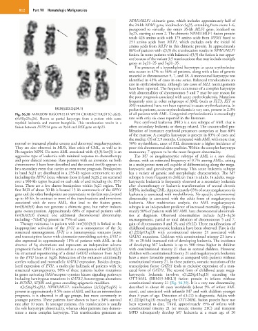
Fig. 56.38 MARROW BASOPHILIA WITH CHARACTERISTIC t(6;9). of all patients with AML. Congenital erythroleukemia is exceedingly
t(6;9)(p23;q24). Shown as partial karyotype from a patient with acute rare with only six cases reported in the literature.
myeloid leukemia and marrow basophilia. This translocation results in a Pure erythroid leukemia (PEL) is a rare subtype of AML that is
fusion between NUP214 gene on 9p34 and DEK gene on 6p23. often secondary leukemia or therapy related. The uncontrolled pro-
liferation of immature erythroid precursors comprises at least 80%
of the marrow. A complex karyotype is present in 83% of cases and
the median OS of 2.9 months. Compared with AML with more than
normal or increased platelet counts and abnormal megakaryopoiesis. 50% erythroblasts, cases of PEL demonstrate a higher incidence of
They are also observed in MDS, blast crisis of CML, as well as in poor-risk chromosomal abnormalities. Within the complex karyotype
Ph-negative MPN. De novo AML associated with t(3;3)/inv(3) is an monosomy 7 appears to be the most frequent abnormality.
aggressive type of leukemia with minimal response to chemotherapy The M7 or megakaryocytic subtype of AML is a rare clonal
and poor clinical outcome. Rare patients with an inversion on both disease, with an estimated frequency of 0.7% among AMLs, arising
chromosome 3 have been described and the second inv(3) appears to in a multipotent stem cell capable of differentiating along the mega-
be a secondary event that carries an even worse prognosis. Breakpoints karyocytic and granulocytic pathway. This acute leukemia subtype
in band 3q21 are distributed in a 235-kb region centromeric to and has a variety of genetic and morphologic characteristics. The M7
including the RPN1 locus, whereas those in band 3q26.2 are scattered subtype is more frequent in children than in adults. In adults, mega-
over a 900-kb region located on each side of and including the EVI1 karyocytic leukemia is frequently observed as a secondary leukemia
locus. There are a few cluster breakpoints within 3q21 region. The after chemotherapy or leukemic transformation of several chronic
first BCR of about 30 kb is located 15 kb centromeric of the RPN1 MPNs, including CML. Approximately 65% of acute megakaryocytic
gene and the other breakpoints are centromeric to the first one, located leukemia is associated with myelofibrosis. No specific chromosomal
up to 60 kb. In contrast to most of the translocations and inversions abnormality is associated with the adult form of megakaryocytic
associated with de novo AML, that lead to the fusion genes, leukemia. After multivariate analysis, the AML megakaryocytic
inv(3)/t(3;3) does not generate a chimeric gene, but rather induces subtype is an independent predictor of increased mortality. Approxi-
gene overexpression. Approximately 68% of reported patients with mately 50% of patients with M7 AML have chromosomal abnormali-
inv(3)/t(3;3) showed one additional chromosomal abnormality, ties at diagnosis. Observed abnormalities include 3q21–3q26
including −7/del(7q) present in 75% of cases. rearrangements, partial or total deletion of chromosomes 5 and 7,
Therapy resistance in patients with inv(3)/t(3;3) is linked to the gain of chromosomes 8 and 19, and t(9;22). Three manifestations of
inappropriate activation of the EVI1 as a consequence of the 3q childhood megakaryocytic leukemia have been observed. First is the
structural rearrangement. EVI1 is a hematopoietic stemness factor t(1;22)(p13;q13) with constitutional trisomy 21 associated with
and transcription factor with chromatin-remodeling activity. EVI1 is GATA1 mutations. Children with constitutional trisomy 21 have a
also expressed in approximately 11% of patients with AML in the 10- to 20-fold increased risk of developing leukemia. The incidence
absence of 3q aberrations and represents an independent adverse of developing M7 leukemia is up to 500 times higher in children
prognostic factor. EVI1 is activated as a consequence of inv(3)/t(3;3) with constitutional trisomy 21 than in normal children. However,
via structural repositioning of a distal GATA2 enhancer from 3q21 children with constitutional trisomy 21 and megakaryocytic leukemia
to the EVI1 locus at 3q26. Relocation of the enhancer additionally have a more favorable prognosis as compared with patients without
confers reduced and monoallelic GATA2 expression. Besides deregu- constitutional trisomy 21. In these patients, somatic mutations of the
lated expression of EVI1, a molecular hallmark of patients with 3q transcription factor GATA1 leads to exclusive expression of a trun-
structural rearrangements, 98% of these patients harbor mutations cated form of GATA1. The second form of childhood acute mega-
in genes activating RAS/receptor tyrosine kinase signaling pathways karyocytic leukemia involves t(1;22)(p13;q13) encoding the
including hemizygous mutations in GATA2, heterozygous alterations OTT-MAL (RBM15-MKL1) fusion protein in infants without
in RUNX1, SF3B1 and genes encoding epigenetic modifiers. constitutional trisomy 21 (Fig. 56.39). It is a very rare abnormality,
t(3;5)(q25;q35), NPM1/MLF1: translocation (3;5)(q25;q35) is described in about 40 cases worldwide (about 5% of infant AML
present in approximately 0.5% cases of AML and has an intermediate cases), and associated with infantile M7 and with children younger
prognosis, it is observed in all age groups, but more commonly in than 3 years of age. Detection of t(1;22) is diagnostic. Adults with
younger patients. These patients have shown to have a 34% survival t(1;22)(p13;q13) encoding the OTT-MAL fusion protein have not
rate after 10 years. In younger patients, this translocation is usually been reported to date. Third, approximately 19% of infants with
the sole karyotypic abnormality, whereas older patients may demon- constitutional trisomy 21 (or mosaic trisomy 21C) and transient
strate a more complex karyotype. This translocation generates an MPD subsequently develop M7 leukemia at a mean age of 20