
Page 930 - Hematology_ Basic Principles and Practice ( PDFDrive )
P. 930
Chapter 56 Conventional and Molecular Cytogenomic Basis of Hematologic Malignancies 813
abnormalities. The overall frequency of MK in AML varies between
6% and 20%. Although in a study of 1058 patients with AML and
abnormal karyotype, 30% had MK (see Fig. 56.27, bottom row).
AML with MK is frequently associated with other adverse risk
cytogenetic abnormalities, such as inv(3), −5 or del(5q), −7 or
del(7q), abnormal (12p), −18/del(18q), abnormal (17p), and a
complex karyotype. The frequency of these recurrent monosomies
are: −7 and −17 in 6%, −18 in 5%, −5 and −21 in 4%, −20 in 3%,
−3, −12, and 22 in 2% and loss of chromosomes 2, 4, 9, 13, and 19
in 1%. MK is a strong prognostic predictor of poor outcome com-
pared with a traditionally defined complex karyotype. Patients with
MK had a 4-year OS of 4% as compared with 21% in patients with
other unfavorable karyotypes but without MK. Responses to induc-
tion therapy and OS of patients who have AML with MK are dismal:
CR rates of 32% and a 4-year survival of 9%. MKs in a study of 248
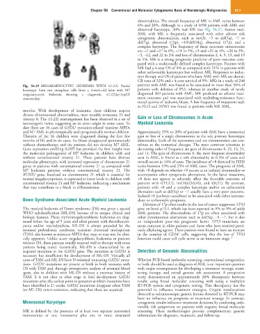
Fig. 56.39 MEGAKARYOCYTIC LEUKEMIA WITH t(1;22). Partial patients with AML was found to be associated in more than 50% of
karyotype from two metaphase cells from a 4-week-old baby with M7 patients with deletion of P53, whereas in another study of newly
megakaryocytic leukemia showing a diagnostic t(1;22)(p13;q13) diagnosed 369 patients with AML, MK predicted an adverse treat-
abnormality. ment outcome and was associated with multidrug-resistance func-
tional activity of leukemic blasts. A low frequency of mutations such
as FLT3 and NPM1 was found in patients with MK AML.
months. With development of leukemia, these children acquire
diverse chromosomal abnormalities, most notably tetrasomy 21 and
trisomy 8. The t(1;22) rearrangement has been observed in a set of Gain or Loss of Chromosomes in Acute
monozygotic twins, suggesting an in utero origin in some cases. To Myeloid Leukemia
date there are 16 cases of GATA1 mutation-related transient MPDs
and M7 AML in phenotypically and cytogenetically normal children. Approximately 15% to 20% of patients with AML have a numerical
Thirteen of the 16 children were diagnosed during the first few gain or loss of a single chromosome as the sole primary karyotypic
months of life and in six cases, the blasts disappeared spontaneously abnormality. Each of the autosomes and sex chromosomes can con-
without chemotherapy and the patients did not develop M7 AML. tribute to the numerical changes. The most common trisomies in
Gene expression profiling (GEP) has provided the first insight into decreasing order of frequency are gain of chromosome 8, 22, 13, 21,
the molecular pathogenesis of M7 leukemia in children with and and 11. The gain of chromosome 8, the most frequent abnormality
without constitutional trisomy 21. These patients have distinct seen in AML, is found as a sole abnormality in 6.3% of cases and
molecular phenotypes, with increased expression of chromosome 21 overall occurs in 16% of cases. The incidence of +8 detected by FISH
genes in patients with constitutional trisomy 21 as compared with varies between 19% and 25% of AML cases. The prognosis of AML
M7 leukemia patients without constitutional trisomy 21. The with +8 depends on whether +8 occurs as an isolated abnormality or
RUNX1 gene, localized on chromosome 21 which is essential for accompanies other cytogenetic aberrations. In the latter situations,
normal megakaryopoiesis, is expressed at lower levels in children with +8 does not appear to adversely affect the favorable outcome of
constitutional trisomy 21 and M7 leukemia, indicating a mechanism patients with t(15;17), inv(16)t(16;16), and t(8;21). By contrast,
that may contribute to a block in differentiation patients with +8 and a complex karyotype and/or an unfavorable
aberration such as del(5q) or −7 usually have a very poor outcome.
Isolated +8 has been considered to be associated with either interme-
Down Syndrome–Associated Acute Myeloid Leukemia diate or unfavorable prognosis.
Deletion of 17p often results in the loss of tumor suppressor TP53
The myeloid leukemia of Down syndrome (DS) was given a special gene on band p13.1, which has been reported in 5% to 9% of adult
WHO subclassification (ML-DS) because of its unique clinical and AML patients. The abnormalities of 17p are often associated with
biologic features. These erythromegakaryoblastic leukemias are diag- other chromosomal aberrations such as del(5q), −5, −7, but is also
nosed before the age of 5 years and often present with thrombocyto- an independent poor-risk prognostic factor. TP53 mutations are
penia and/or myelodysplasia. ML-DS is always preceded by the more common in older patients and those who have received previ-
neonatal preleukemic syndrome, transient abnormal myelopoiesis ously alkylating agents. These patients were found to have an increase
+
(TAM; also known as transient MPD) that may, or may not, be clini- in the number of CD34 cells, suggesting that the loss of TP53
cally apparent. Unlike acute megakaryoblastic leukemias in patients function could cause cell cycle arrest at an immature stage. 17
without DS, these patients usually respond well to therapy with most
patients being cured. Genetically, ML-DS is characterized by an
acquired mutation in the GATA1 gene. The mutation in GATA1 is Detection of Genomic Abnormalities
necessary but insufficient for development of ML-DS. Virtually all
cases of TAM and ML-DS have N-terminal truncating GATA1 muta- Whether PCR-based molecular screening, conventional cytogenetics,
tions. GATA1 mutations are present at birth in both neonates with or both, should be used at diagnosis of AML is an important question
DS with TAM and, through retrospective analysis of neonatal blood with major consequences for developing a treatment strategy, moni-
spots, also in children with ML-DS without a previous history of toring therapy, and overall genetic risk assessment. A prospective
TAM. It is not clear at what stage in fetal development GATA1 study demonstrated an approximately 20% discrepancy between
mutations arise; the earliest point in gestation at which mutations have results using broad molecular screening with using a multiplex
been identified is 21 weeks. GATA1 mutations disappear when TAM RT-PCR system and cytogenetic testing. This discrepancy has the
(or ML-DS) enters remission, indicating that these are acquired. potential to influence treatment strategies. Cryptic translocations
detected as submicroscopic genetic lesions detected by RT-PCR may
have no influence on prognosis or treatment strategy. In contrast,
Monosomal Karyotype cytogenetic results influence treatment decisions by conferring unfa-
vorable risk assignment on patients with negative broad molecular
MK is defined by the presence of at least two separate autosomal screening. These methodologies provide complementary genetic
monosomies or one monosomy plus one or more structural information for diagnosis, treatment, and follow-up.