
Page 1077 - Williams Hematology ( PDFDrive )
P. 1077
1052 Part VIII: Monocytes and Macrophages Chapter 67: Structure, Receptors, and Functions of Monocytes and Macrophages 1053
26
The most constant and characteristic ultrastructural features of differentiation, activation, recognition, migration, and function
macrophages are the electron-dense membrane-bound lysosomes that of the monocyte/macrophage. Monocytes have been classified into
often can be seen fusing with phagosomes to form secondary lysosomes. distinct subtypes based on surface expression of CD14 and CD16,
Within the secondary lysosomes, ingested cellular, bacterial, and non- molecules that form part of the lipopolysaccharide (LPS) toll-like
cellular material can be seen in various stages of degradation, often receptor (TLR) and one of the immunoglobulin FcRs, respectively.
recognizable as degenerating mitochondria or nuclear material. These These include CD14+-bright/CD16– monocytes, CD14+-dim/CD16+
secondary lysosomes also contain partially degraded material from the monocytes, and CD14-dim/CD16+ monocytes. Monocyte hetero-
late stages of the endocytic process, often appearing as multilamellar geneity was initially divided into the CD14+-bright/CD16-negative
lipid bodies. Microtubules and microfilaments are prominent in macro- cells, which comprise 90 to 95 percent of total circulating monocytes
27
phages. Actin- and myosin-like proteins have been isolated from mono- (classical monocyte) —CD14-bright or dim refer to the fluorescence
cytes and partially characterized. Resting macrophages have irregular magnitude of staining using a specific CD14 monoclonal antibody.
cell borders and pseudopodia pushed out in all directions. Their cyto- The minor subset is CD14-dim, CD16-positive, and less phagocytic
plasm has rough endoplasmic reticulum and Golgi complex in the per- than the classical monocyte. The classical monocyte produces reactive
inuclear area. Lipid globules, primary lysosomes, and mitochondria are oxygen species (ROS) and cytokines in response to TLR engagement.
characteristically prominent. Activated monocytes/macrophages are The minor subset selectively secretes tumor necrosis factor (TNF)-α,
motile cells that extend a leading pseudopod as they move forward. 25 IL-13, and CCL2 in response to viruses and immune complexes con-
taining nucleic acids via TLR-7, TLR-8, MyD88-MEK (myeloid dif-
28
ferentiation factor 88–MAPK kinase), and AHD. This minor subset,
RECEPTORS CD14-dim, is competent in (SR [scavenger receptor]) function of
29
vascular, intraluminal debris and uptake of immune complexes.
30
MEMBRANE RECEPTORS AND OTHER In addition, their phenotype is related to the ability to produce and
SURFACE PROTEINS OF MONOCYTES secrete select cytokines. 31
Macrophages are proficient at endocytosis (both fluid phase and
AND MACROPHAGES receptor-mediated) and are highly professional phagocytes of particu-
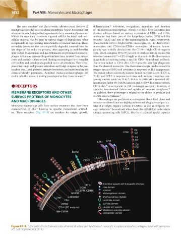
Monocyte/macrophage cells have surface receptors that have been lates of all origin, organic (cellular, microbial) as well as inorganic for-
characterized by their binding to specific monoclonal antibod- eign materials. In contrast, when dendritic cells (DCs) mature into
32
ies. These receptors (Fig. 67–8) are markers for origin, growth, antigen-presenting cells (APCs), they have reduced uptake capacity
C2
CD31 C2
C2
LFA1 CR3 C2
C2
C2
EMR2
CR1
CX 3
CCR2
CD36
α2
α1
β2
β1
v
v c CD14 N-terminal repeats with β-propeller structure
c CD86 I-like domain
M-CSFR (CD115) I-domain
MHC11 Immunoglobulin domain
β ITAM L-selection Short concensus repeats
α CD4 Lectin-like domain
CD33 EGF-like domain
CD16 (FC receptor) Leucine-rich repeats
Membrane-spanning domain
GM-CSF-R
Hemopoeitin domain
Figure 67–8. Schematic of selected molecules of varied structure and functions of monocyte receptors and surface antigens. (Used with permission
of S. Seif, GraphisMedica, 2014.)
Kaushansky_chapter 67_p1043-1074.indd 1052 9/21/15 10:43 AM