
Page 1095 - Williams Hematology ( PDFDrive )
P. 1095
1070 Part VIII: Monocytes and Macrophages Chapter 67: Structure, Receptors, and Functions of Monocytes and Macrophages 1071
TABLE 67–7. Immunomodulation of Macrophage Phenotype
Stimulus Category Markers Function
Microbial (bacterial) Innate activation Induction of MARCO Enhanced phagocytosis
Costimulatory molecules Antigen presentation
CD200 Inhibition (CD200R)
IFN-γ Classical activation Induction MHC II Cell-mediated immunity/delayed-type
hypersensitivity
Potentiation innate markers
- TNF-α Proinflammatory
- iNOS induction Antimicrobial (NO) signaling
- NADPH, respiratory burst Host defense, inflammation
LGP47 induction Association with phagosome/intracellu-
lar pathogen killing
Downregulation of MR Unknown
Modulation of FcR expression
Proteasomal composition Antigen presentation
IL-4/IL-13 Alternative Enhanced MR Endocytosis
activation
Induction arginase Humoral immunity
Induction YM1, FIZZ1 (mouse) Th2-responses, allergy, antiparasitic
Induction CCL17 (MDC) and CCL22 (TARC) Immunity, repair/fibrosis
Fusion, giant cell formation
Upregulation CD23 (FcRε)
Immune complexes Modified activation Selective IL-12 downregulation, IL-10 induction
IL-10 Deactivation Downregulation MHC II
TGF-β Deactivation Downregulation of proinflammatory NO and ROI
Glucocorticoids Deactivation CD163 induction, monocyte recruitment down- Antiinflammatory
regulated, ACE induction, Stabilin induction
Homeostatic clearance of hemoglobin/
haptoglobin complexes
IFN, interferon; IL, interleukin; iNOS, inducible nitric oxide synthase; MARCO, macrophage receptor with collagenous structure; MDC, mac-
rophage-derived chemokine; MHC, major histocompatibility complex; MR, mannose receptor; NADPH, nicotinamide adenine dinucleotide
phosphate; NO, nitric oxide; ROI, reactive oxygen intermediate; TARC, thymus and activation-regulated chemokine; TGF, transforming growth
factor; TNF, tumor necrosis factor.
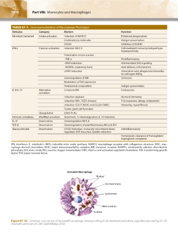
Activated Macrophage
“Ruffles”
Cell membrane
Lysosomes
Mitochondrion
Nucleus
Figure 67–22. Schematic cross-section of “activated” macrophage, showing ruffling of cell membrane and cellular organelles (also see Fig. 67–15).
(Used with permission of S. Seif, GraphisMedica, 2014.)
Kaushansky_chapter 67_p1043-1074.indd 1070 9/21/15 10:44 AM