
Page 1151 - Williams Hematology ( PDFDrive )
P. 1151
1126 Part VIII: Monocytes and Macrophages Chapter 72: Gaucher Disease and Related Lysosomal Storage Diseases 1127
accumulated lipid on platelet membranes. Factor XI deficiency is com- Other biomarkers correlate better with the extent of glucocerebroside
92
mon among Ashkenazi Jewish patients because of its high coincidental storage. The most widely used biomarker is chitotriosidase, which
98
prevalence in this ethnic group. 93 is undetectable in healthy subjects (its physiologic role is unknown),
Bleeding tendency may also result from defective aggregation or but is elevated, often several thousand-fold, in patients with Gaucher
adhesion of platelets and therefore platelet function and/or thromboe- disease. Chitotriosidase measurement is useful for monitoring both
33
lastography should be tested before surgical and dental procedures and untreated patients, to assess stability versus deterioration, and treated
labor. 44,94 patients, to assess response to therapy. A change in chitotriosidase levels
rather than absolute values is used for monitoring. In approximately 6
Biochemical and Immunologic Findings percent of people, it is undetectable, and for those patients, measure-
In most patients, liver function tests are within normal limits but in con- ments of chemokine CCL18/PARC which is predominantly produced
junction with more severe disease, splenectomy, and/or comorbidities by Gaucher cells, can be used. 99
(hepatitic B and/or C, or autoimmune diseases) abnormal liver function A potentially more sensitive and more specific biomarker has been
tests may be seen. Because of the increased prevalence of cholelithiasis, 95,96 identified: the lyso-glucosylsphingosine (lyso-Gb1), which may be
100
cholestatic findings may occur. Renal function tests are usually normal. 64 preferred as a more reproducible biomarker, using a more operator-
Many patients present with polyclonal gammopathies. Monoclonal friendly assay.
gammopathies are found in 1 to 20 percent of patients, particularly older Serum iron levels may be low in patients because of iron deficiency
patients. 79–82 Increased levels of autoantibodies have been reported, and related to bleeding or chronic inflammation. Deficiencies of vitamin B 12 101
97
may indicate coincide with autoimmune diseases such as Hashimoto and vitamin D have been described, albeit these are also very common
102
thyroiditis, rheumatoid arthritis, or immune hemolytic anemia. in the general population. Serum ferritin levels are usually elevated.
Biochemical abnormalities have been used as surrogate mark-
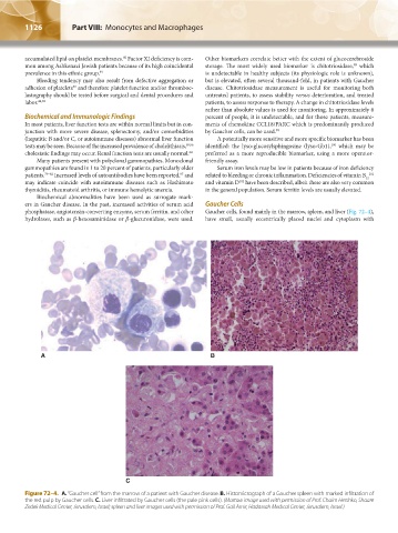
ers in Gaucher disease. In the past, increased activities of serum acid Gaucher Cells
phosphatase, angiotensin-converting enzyme, serum ferritin, and other Gaucher cells, found mainly in the marrow, spleen, and liver (Fig. 72–4),
hydrolases, such as β-hexosaminidase or β-glucuronidase, were used. have small, usually eccentrically placed nuclei and cytoplasm with
A B
C
Figure 72–4. A. “Gaucher cell” from the marrow of a patient with Gaucher disease. B. Histomicrograph of a Gaucher spleen with marked infiltration of
the red pulp by Gaucher cells. C. Liver infiltrated by Gaucher cells (the pale pink cells). (Marrow image used with permission of Prof. Chaim Hershko, Shaare
Zedek Medical Center, Jerusalem, Israel; spleen and liver images used with permission of Prof. Gail Amir, Hadassah Medical Center, Jerusalem, Israel.)
Kaushansky_chapter 72_p1121-1134.indd 1126 9/17/15 3:53 PM