
Page 1614 - Williams Hematology ( PDFDrive )
P. 1614
1588 Part XI: Malignant Lymphoid Diseases Chapter 96: Pathology of Lymphomas 1589
precursor lymphoid neoplasms, mature B-cell neoplasms, mature T-
and NK-cell neoplasms, and Hodgkin lymphoma.
Distinctive lymphoma entities were identified based upon a combi-
nation of morphologic, immunophenotypic, genetic, and clinical features.
The 2008 WHO classification includes several provisional entities and cat-
egories of unclassifiable neoplasms with features intermediate between two
distinct entities. This allows the classification to retain flexibility so that new
data that further identify distinct diseases within these entities can be incor-
porated. In distinction from the Working Formulation, lymphomas were
not classified based on clinical outcome. The WHO classification agreed
that each type of lymphoma that was identified by pathologic and clinical
features could have a spectrum of clinical aggressiveness, and that lump-
ing distinct entities into groups based on clinical outcome would inhibit
the development of targeted therapeutic approaches. Therefore, the WHO
classification represents a complete change from the Working Formulation,
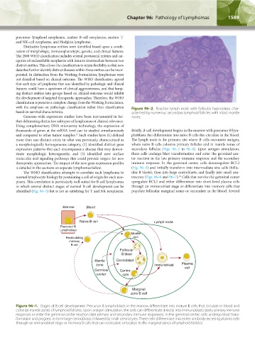
with the emphasis on pathologic classification rather than classification Figure 96–2. Reactive lymph node with follicular hyperplasia, char-
based on survival characteristics. acterized by numerous secondary lymphoid follicles with intact mantle
Genome-wide expression studies have been instrumental in fur- zones.
ther delineating distinctive subtypes of lymphomas of clinical relevance.
Using complementary DNA microarray technology, the expression of
thousands of genes at the mRNA level can be studied simultaneously Briefly, B-cell development begins in the marrow with precursor B lym-
15
and compared to other tumor samples. Such studies have (1) defined phoblasts that differentiate into naïve B cells that circulate in the blood.
more than one distinct entity in what was previously characterized as The lymph node is the primary site where B cells encounter antigen,
a morphologically homogeneous category, (2) identified distinct gene where naïve B cells colonize primary follicles and in mantle zones of
expression patterns that each encompasses a disease that may demon- secondary follicles (Figs. 96–2 to 96–4). Upon antigen stimulation,
strate morphologic heterogeneity, and (3) identified new surface these cells undergo blast transformation and enter the germinal cen-
molecules and signaling pathways that could provide targets for new ter reaction in the late primary immune response and the secondary
therapeutic approaches. The impact of the new gene expression profiles immune response. In the germinal center, cells downregulate BCL2
is detailed in the sections on separate lymphomas below. (Fig. 96–5) and initially transform into intermediate-size cells (follic-
The WHO classification attempts to correlate each lymphoma to ular B blasts), then into large centroblasts, and finally into small cen-
16
normal lymphocyte biology by postulating a cell of origin for each neo- trocytes (Figs. 96–6 and 96–7). Cells that survive the germinal center
plasm. This correlation is particularly well-suited for B-cell lymphomas upregulate BCL2 and either differentiate into short-lived plasma cells
in which several distinct stages of normal B-cell development can be through an immunoblast stage or differentiate into memory cells that
identified (Fig. 96–1) but is not as satisfying for T and NK neoplasms. populate follicular marginal zones or recirculate in the blood. Several
Marrow Blood
Naïve B cell Lymph node
Precursor B
Lymphoblast
Mantle
Recirculating cell
memory
B cell
Follicular
B blast
Mantle Centroblast
zone Immunoblast Plasma
Germinal Centro- cell
center cyte
Marginal
zone B cell
Figure 96–1. Stages of B-cell development. Precursor B lymphoblasts in the marrow differentiate into mature B cells that circulate in blood and
colonize mantle zones of lymphoid follicles. Upon antigen stimulation, the cells can differentiate directly into immunoblasts (early primary immune
response) or enter the germinal center reaction (late primary and secondary immune responses). In the germinal center, cells undergo blast trans-
formation and progress to form large centroblasts, followed by small centrocytes. These cells differentiate into either antibody-secreting plasma cells
through an immunoblast stage or memory B cells that can recirculate or localize to the marginal zones of lymphoid follicles.
Kaushansky_chapter 96_p1587-1602.indd 1589 9/18/15 6:06 PM