
Page 1619 - Williams Hematology ( PDFDrive )
P. 1619
1594 Part XI: Malignant Lymphoid Diseases Chapter 96: Pathology of Lymphomas 1595
cyclin D1–negative cases have chromosomal translocations involving
30
the cyclin D2 gene. SOX11 expression is a highly specific immuno-
histochemical marker for mantle cell lymphoma, and can identify cases
31
that are negative for cyclin D1. Overall, patients with mantle cell
lymphoma have a median survival of approximately 3 years, but gene
expression data that determine tumor cell proliferation are able to iden-
29
tify patient subsets that differ in median survival by more than 5 years
(Chap. 100).
Cyclin D1–positive lymphocytes in mantle zones can be an inci-
dental finding in reactive lymphoid follicles, a condition referred to as
in situ mantle cell lymphoma. These appear to have an indolent behavior
and do not require treatment. 32
FOLLICULAR LYMPHOMA
Follicular lymphoma is a proliferation of cells that correspond to nor-
mal germinal center cells, retaining expression of germinal center Figure 96–18. Center of a neoplastic follicle in grade 1 follicular lym-
33
markers (BCL6, CD10), and demonstrates a follicular architecture phoma with almost exclusively small centrocytes.
(Fig. 96–17) imparted by nodular aggregates of CD21-positive follic-
ular dendritic cells. Follicular lymphomas are composed of a variable
mixture of centrocytes (small cleaved cells) and centroblasts (large non-
cleaved cells). They can be divided into three grades (grades 1 to 3) based
on the number of centroblasts present. The most common is grade 1
(0–5 centroblasts per high-power microscopic field), previously known
as follicular small-cleaved-cell lymphoma (Fig. 96–18). Both grade 1
and grade 2 tumors are indolent, and distinguishing between them is
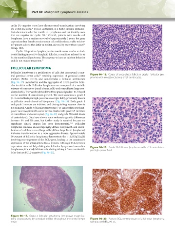
not required. Grade 3 follicular lymphoma (>15 centroblasts per high-
power microscopic field) can be further divided into grade 3A (mixture
of centroblasts and centrocytes) (Fig. 96–19) and grade 3B (solid sheets
of centroblasts). Data have shown some molecular genetic differences
between 3A and 3B cases, but further study is required because no
significant clinical impact has been demonstrated. 34,35 Follicular
lymphoma can have an accompanying diffuse component, and identi-
fication of a diffuse area of large cells (diffuse large B-cell lymphoma)
indicates transformation to a more aggressive disease. Approximately
90 percent of follicular lymphoma demonstrate the t(14;18)(q32;q21)
involving rearrangement of the BCL2 gene, leading to the constitutive
expression of the antiapoptotic BCL2 protein. Although BCL2 protein
expression does not help distinguish follicular lymphoma from other Figure 96–19. Grade 3A follicular lymphoma with >15 centroblasts
lymphomas, it is a helpful feature in distinguishing it from reactive fol- per high-power field.
licles that are BCL2-negative (Fig. 96–20).
Figure 96–17. Grade 2 follicular lymphoma (low-power magnifica-
tion), characterized by crowded follicles throughout the entire lymph Figure 96–20. Positive BCL2 immunostain of a follicular lymphoma
node. (contrast with Fig. 96–5).
Kaushansky_chapter 96_p1587-1602.indd 1594 9/18/15 6:07 PM