
Page 1618 - Williams Hematology ( PDFDrive )
P. 1618
1592 Part XI: Malignant Lymphoid Diseases Chapter 96: Pathology of Lymphomas 1593
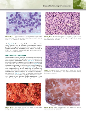
Figure 96–12. Imprint preparation of lymphoplasmacytic lymphoma Figure 96–14. Mantle cell lymphoma with a diffuse pattern, charac-
demonstrating small lymphocytes and cells with plasmacytoid features terized by a monomorphous infiltrate of small irregular lymphocytes
(eccentric nuclei and bluish cytoplasm). with numerous mitotic figures.
cells (Fig. 96–12). These cases typically do not express CD5, less often
involve blood, and often are associated with a monoclonal immuno-
globulin M serum protein that can cause hyperviscosity or cryoglob-
ulinemia (Waldenström macroglobulinemia). Somatic mutations in
MYD88 are a commonly recurring and highly specific feature of Wal-
denström macroglobulinemia. 26
MANTLE CELL LYMPHOMA
Mantle cell lymphoma most commonly involves lymph nodes, but it can
involve extranodal sites, including the gastrointestinal tract, as a clinical
variant known as lymphomatous polyposis (Fig. 96–13). It typically is
composed of a uniform population of small lymphocytes with cleaved
nuclei and a virtual absence of large transformed cells (Fig. 96–14). 27,28
It most commonly has a diffuse growth pattern, but it can show a nod-
ular or, more rarely, a mantle zone pattern (Fig. 96–15). The postulated
cell of origin is the B cell of the inner mantle zone. The lymphoma cells
coexpress CD5, as does chronic lymphocytic leukemia, but mantle cell Figure 96–15. Mantle cell lymphoma with a mantle-zone pattern,
lymphoma can be distinguished by lack of CD23 expression and expres- characterized by monomorphous small lymphocytes surrounding a
sion of cyclin D1 (Fig. 96–16). Cyclin D1 expression results from the benign germinal center.
chromosomal translocation t(11;14)(q13;q32) characteristic of man-
tle cell lymphoma. Gene expression data have demonstrated a subset
of mantle cell lymphomas that are cyclin D1–negative. Some of these
29
Figure 96–13. Large bowel involved with mantle cell lymphoma Figure 96–16. Mantle cell lymphoma with mantle-zone pattern
(multiple lymphomatous polyposis). stained with antibody to cyclin D1.
Kaushansky_chapter 96_p1587-1602.indd 1593 9/18/15 6:07 PM